Monday, June 14, 2010

LEARNING RADIOLOGY


Pneumothorax

  • Presence of air in the pleural space
    • Anatomy
      • Visceral pleura is adherent to lung surface
      • There is no air in the pleural space normally
      • The introduction of air into the pleural space separates the visceral from the parietal pleura
        • In contradistinction, the visceral and parietal pleura usually do not separate from each other in obstructive atelectasis
  • Pathophysiology
    • Either from disruption of visceral pleura
    • Or, trauma to parietal pleura
  • Clinical findings
    • Acute onset of
      • Pleuritic chest pain
      • Dyspnea (in 80-90%)
      • Cough
      • Back or shoulder pain
  • Etiologies
    • Penetrating trauma
    • Blunt trauma
      • May be due to rib fracture
      • May be caused by increased intrathoracic pressure
      • May lead to bronchial rupture
        • “Fallen lung sign” (ptotic lung sign) --  hilum of lung is below expected level within chest cavity
        • Persistent pneumothorax with functioning chest tube
    • Iatrogenic
      • Tracheostomy
      • Central venous catheter attempt or insertion
      • Mechanical ventilation
        • May occur in up to 25% of patients maintained on PEEP
        • May be bilateral or under tension
      • Thoracic irradiation
    • Spontaneous pneumothorax
      • Most common etiology
      • Cause
        • Rupture of subpleural blebs in apical region of lung
      • Age
        • 20-40 years
        • M:F = 8:1
        • Especially in patients who are tall and thin
        • Smokers
      • Prognosis
        • Recurrence in 30% on same side
        • Recurrence in 10% on contralateral side
      • Treatment
        • Simple aspiration (success in >50%)
        • Tube thoracostomy (effective in 90%)
    • Other causes of a pneumothorax
      • Neonatal disease
        • Meconium aspiration
        • Respirator therapy for hyaline membrane disease
      • Malignancy
        • Primary lung cancer
        • Lung metastases, especially from osteosarcoma
          • Also pancreas, adrenal, Wilms tumor
      • Pulmonary infections
        • Tuberculosis
        • Necrotizing pneumonia
        • Coccidioidomycosis
        • Hydatid disease
        • Pertussis
        • Acute bacterial pneumonia
        • Staphylococcal septicemia
        • AIDS (Pneumocystis carinii, Mycobacterium tuberculosis, atypical mycobacteria)
      • Complication of pulmonary fibrosis
        • Histiocytosis X
        • Idiopathic
        • Cystic fibrosis
        • Sarcoidosis
        • Scleroderma
        • Eosinophilic granuloma
        • Interstitial pneumonitis
        • Rheumatoid lung
        • Idiopathic pulmonary hemosiderosis
        • Pulmonary alveolar proteinosis
        • Biliary cirrhosis
      • Asthma or emphysema
        • Produce a second peak incidence of pneumothorax from 45-65 years of age
        • Due to rupture of peripheral emphysematous areas
      • “Catamenial pneumothorax” is a recurrent spontaneous pneumothorax that occurs during menstruation and is associated with endometriosis of the diaphragm
        • R >> L
      • Marfan’s syndrome
      • Ehlers-Danlos syndrome
      • Pulmonary infarction
      • Lymphangiomyomatosis and tuberous sclerosis
        • Incidence of pneumothorax is particularly high in lymphangiomyomatosis and histiocytosis X
  • Types of pneumothorax
    • Closed pneumothorax = intact thoracic cage
    • Open pneumothorax = "sucking" chest wound
    • Tension pneumothorax
      • Accumulation of air within pleural space due to free ingress and limited egress of air
      • Pathophysiology:
        • Intrapleural pressure exceeds atmospheric pressure in lung during expiration (check-valve mechanism)
      • Frequency
        • In 3-5% of patients with spontaneous pneumothorax
        • Higher in barotrauma (mechanical ventilation)
    • Simple pneumothorax –no shift of the heart or mediastinal structures
  • Imaging findings in pneumothorax
    • Must see the visceral pleural white line
      • Very thin white line that differs from a skin fold by its thickness
    • Absence of lung markings distal or peripheral to the visceral pleural white line
      • Not evidence enough to say there is a pneumothorax only if there are no lung markings seen
        • No lung markings will be seen with bullous disease
        • Bullae have a concave surface facing the chest wall
          • Pneumothorax almost always has a convex surface facing the chest wall
    • Displacement of mediastinum and/or anterior junction line
    • Deep sulcus sign
      • On frontal view, larger lateral costodiaphragmatic recess than on opposite side
      • Diaphragm may be inverted on side with deep sulcus
    Total / subtotal lung collapse
    • This is passive or compressive atelectasi
  • Collapse of SVC or IVC due to decreased systemic venous return and decreased cardiac output
  • Tension hydropneumothorax
    • Sharp delineation of visceral pleural by dense pleural space
    • Mediastinal shift to opposite side
    • Air-fluid level in pleural space on erect chest radiograph
  • Radiographic signs in upright position
    • White margin of visceral pleura separated from parietal pleura
    • Usually seen in the apex of the lung
    • Absence of vascular markings beyond visceral pleural margin
    • May be accentuated by an expiratory film in which lung volume is reduced while amount of air in pneumothorax remains constant so that relative size of pneumothorax appears to increase
  • Radiographic signs in supine position
    • Anteromedial pneumothorax (earliest location)
    • Outline of medial diaphragm under cardiac silhouette
    • Deep sulcus sign
      • Decubitus views of the chest may demonstrate a pneumothorax on the side that is non-dependent
        • Left lateral decubitus view for right-sided pneumothorax
        • Right lateral decubitus view for left-sided pneumothorax
  • Subpulmonic pneumothorax (second most common location)
    • Hyperlucent upper abdominal quadrant
    • Deep lateral costophrenic sulcus
    • Sharply outlined diaphragm in spite of parenchymal disease
    • Visualization of anterior costophrenic sulcus
    • Visualization of inferior surface of lung
  • Apicolateral pneumothorax (least common location)
    • Visualization of visceral pleural line
  • Posteromedial pneumothorax (in presence of lower lobe collapse)
    • Lucent triangle with vertex at hilum
    • V-shaped base delineating costovertebral sulcus
    • Pneumothorax outlines pulmonary ligament
  • Pitfalls in diagnosis
    • Skin fold
      • Thicker than the thin visceral pleural white line
    • Air trapped between chest wall and arm
      • Will be seen as a lucency rather than a visceral pleural white line
    • Edge of scapula
      • Follow contour of scapula to make sure it does not project over chest
    • Overlying sheets
      • Usually will extend beyond the confines of the lung
    • Hair braids
  • Prognosis
    • Resorption of pneumothorax occurs at a rate of 1.25% per day (accelerated by increasing inspired oxygen concentrations)
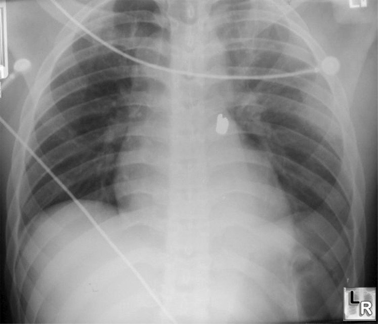
Left pneumothorax-deep sulcus sign., Lucency at left costophrenic angle which projects well below the
costophrenic angle on the opposite side is the "Deep sulcus sign" indicating the
presence of a pneumothorax on a supine radiograph of the chest
(Bullet is seen overlying the heart)

    
Thoracic Radiology: The Requisites
Dahnert 4th Edition

No comments:

Post a Comment