Ankle radiograph
AP projection.
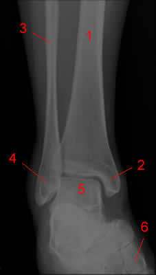 1, Tibia.
2, Medial Malleolus.
3, Fibula.
4, Lateral Malleolus.
5, Talus.
6, 1st Metatarsal Bone.
Lateral projection.
 1, Cuneiform Bone.
2, Navicular Bone.
3, Talus.
4, Tibia.
5, Fibula.
6, Calcaneus.
7, Cuboid Bone.
Knee radiograph
Knee Radiograph - AP
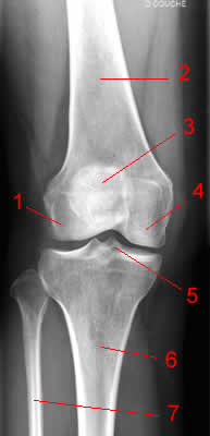 1, Lateral condyle of femur.
2, Femur.
3, Patella.
4, Medial condyle of femur.
5, Medial intercondylar tubercle of tibia.
6, Tibia.
7, Fibula.
Knee Radiograph - Lateral
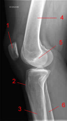 1, Patella.
2, Tuberosity of tibia.
3, Tibia.
4, Femur.
5, Medial condyle of femur.
6, Fibula.
Hip radiography
Hip radiography, AP view.
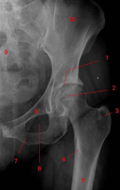
1, Acetabular fossa.
2, Head femoral.
3, Greater trochanter.
4, Lesser trochanter.
5, Femur.
6, Obturator foramen.
7, Inferior pubic ramus.
8, Superior pubic ramus.
9, Sacrum.
10, Iliac wing.
Hip radiography, "frog leg" lateral view.
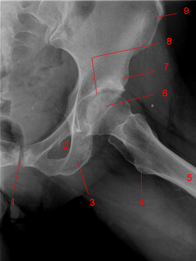
1, Symphysis pubis.
2, Obturator foramen.
3, Ischium.
4, Lesser trochanter.
5, Femur.
6, Femoral head.
7, Anterior inferior iliac spine.
8, Acetabular fossa.
9, Anterior superior iliac spine.
Pelvis Radiograph
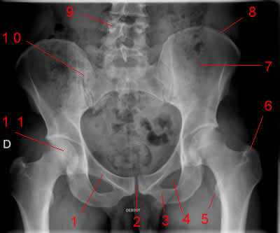 1 Superior Ramus of Right Pubis
2 Symphysis Pubis
3 Inferior Ramus of Left Pubis
4 Left obturator foramen
5 Left lesser Trochanter
6 Left Greater Trochanter
7 Left iliac wing
8 Iliac crest
9 Vertebral Pedicle (Lumbar Spine)
10 Right Sacro-iliac joint
11 Head of right femur
Foot X-ray
Foot X-ray AP
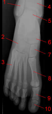 1, Fibula.
2, Cuboid.
3, 5th metatarsal.
4, Tibia.
5, Talus.
6, Navicular.
7, Cuneiform
8, 1st metatarsal
9, proximal phalanx
10, distal phalanx
Foot X-ray oblique
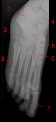 1, Calcaneus.
2, Cuboid.
3, 5th metatarsal.
4, Talus.
5, Navicular.
6, Cuneiform.
7, Interphalangeal joint.
Abdominal X-ray
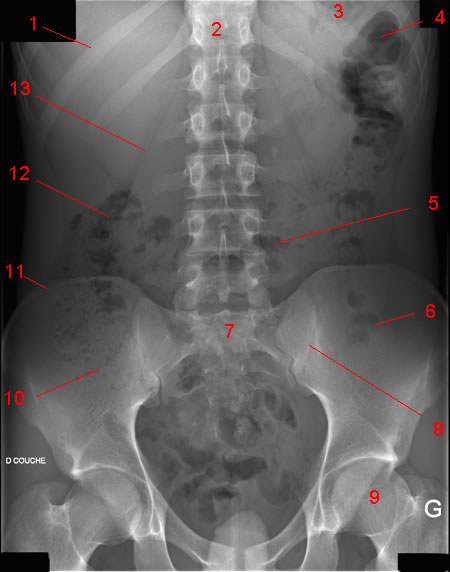
1, 11th rib.
2, Vertebral body (TH 12).
3, Gas in stomach.
4, Gas in colon (splenic flexure).
5, Gas in transverse colon.
6, Gas in sigmoid.
7, Sacrum.
8, Sacroiliac joint.
9, Femoral head.
10, Gas in cecum
11, Iliac crest.
12, Gas in colon (hepatic flexure).
13, Psoas margin.
Elbow Radiograph
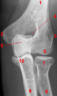 Elbow Radiograph - AP projection1, Humerus. 2, Medial epicondyle. 3, Lateral epicondyle. 4, Olecranon fossa. 5, capitellum. 6, Radius. 7, Radial Head.8, Ulna. 9, Olecranon process. 10, Coronoid process. 11, Anterior fat pad (here: fat pad sign).
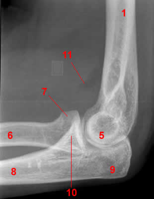 Elbow Radiograph - Lateral projection
Elbow Radiograph - AP projection
Elbow Radiograph - Lateral projection


Wrist radiographs. Image 1 | ||||||
 | ||||||
Previous | Next 1, Ulnar styloid process. 2, Lunate. 3, Radius. 4, Navicular.5, Trapezium (Greater multangular). 6, 1st Metacarpal. 7, Trapezoid (Lesser multangular). 8, Capitate. 9, Hamate. 10, Triquetrum. 11, Pisiform. .
|
Lumbar Spine X-ray
Lumbar spine X-ray, AP projection
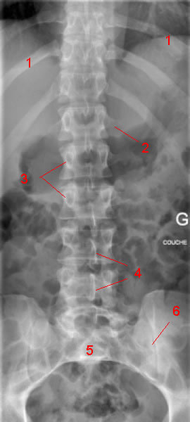 1, rib.
2, Transverse process.
3, Pedicle.
4, Spinous Process.
5, Sacrum.
6, Sacroiliac joint.
Lumbar spine X-ray, lateral view
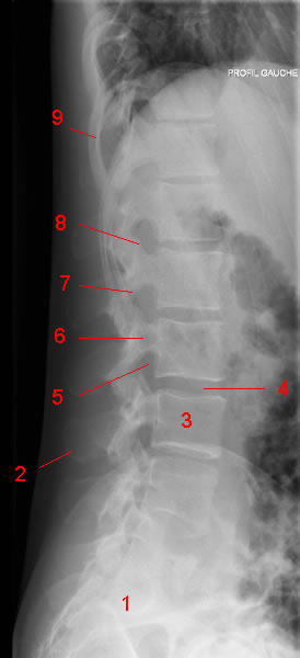 1, Sacrum.
2, Spinous Process.
3, Vertebral body.
4, Intervertebral disc space.
5, Intervertebral foramina.
6, Pedicle.
7, Inferior articulating facet.
8, Superior articulating facet.
9, Rib .
1, rib.2, Transverse process.
3, Pedicle.
4, Spinous Process.
5, Sacrum.
6, Sacroiliac joint.
1, Sacrum.2, Spinous Process.
3, Vertebral body.
4, Intervertebral disc space.
5, Intervertebral foramina.
6, Pedicle.
7, Inferior articulating facet.
8, Superior articulating facet.
9, Rib .
Hand radiography
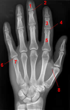 Hand X-ray - AP1, Distal phalanx. 2, Distal interphalangeal joint. 3, Middle phalanx. 4, Proximal interphalangeal joint. 5, Proximal phalanx. 6, Metacarpophalangeal joint. 7, Head of 5th metacarpal. 8, Sesamoid bone.
Hand X-ray - AP
Forearm X-ray
AP Projection.
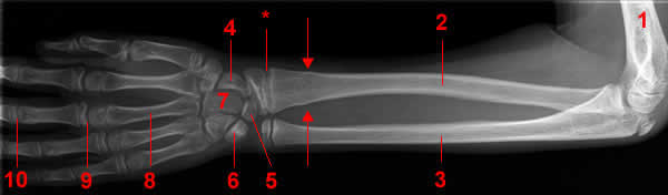 1, Humerus. 2, Radius. 3, Ulna. 4 Navicular Bone 5 Lunate Bone. 6 Triquetrum. 7 Capitatum bone. 8, Metacarpal bone. 9 Metacarpophalangeal joint. 10 Proximal phalangeal joint. 11 Proximal Phalanx. 12 Distal phalanx. Arrow,Fracture.
*, Epiphysial plate.Lateral projection.
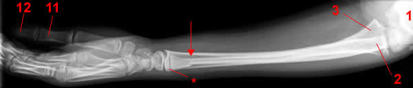
*, Epiphysial plate.
Shoulder X-ray
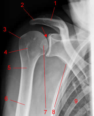
Shoulder X-ray, AP projection
1, Clavicle. 2, Acromion. 3, Greater tubercle. 4, Lesser tubercle. 5, Neck of Humerus. 6, Humerus. 7, Coracoid Process. 8, Axillary border of scapula. 9, Rib.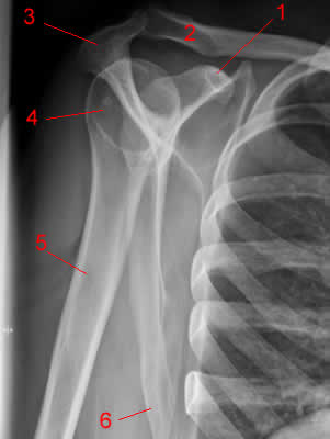
Shoulder X-ray: lateral view
1, Coracoid Process. 2, Clavicle. 3, Acromion. 4, Head of Humerus. 5, Humerus. 6, Axillary border of scapula.
Thoracic spine X-ray. Image 4 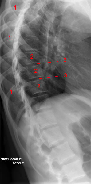

Thoracic spine X-ray. Image 3 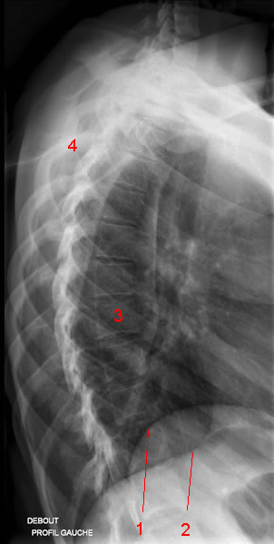
Thoracic spine X-ray. Image 2 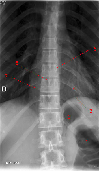
Thoracic spine X-ray. Image 1 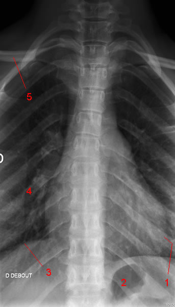
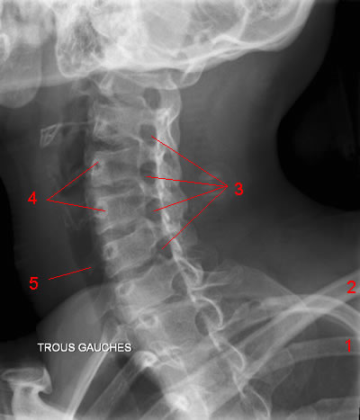
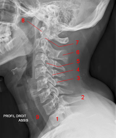
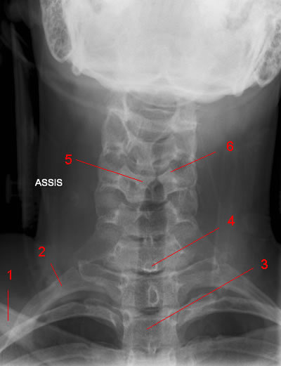
Cervical Spine X-Ray: Image 5 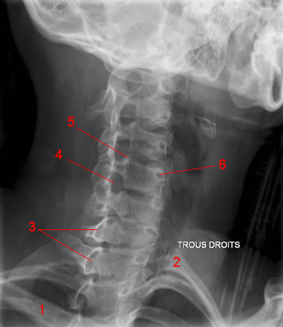
Thoracic spine X-ray. Image 4 |
|
Thoracic spine X-ray. Image 3 |
|
Thoracic spine X-ray. Image 2
Thoracic spine X-ray. Image 1
Cervical Spine X-Ray: Image 5
Cervical Spine X-Ray: Image 3 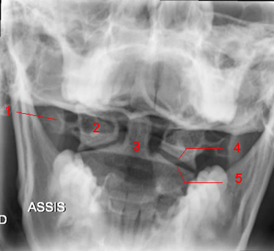
Cervical Spine X-Ray: Image 3
Cervical Spine X-Ray: Image 2 |
 |
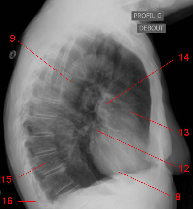
Chest X-ray
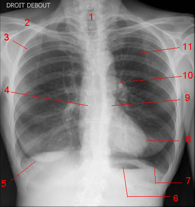
AP Projection.
1, Trachea. 2, Clavicle. 3, 4th posterior rib. 4, Right main bronchus. 5, Right breast shadow. 6, Gastric air bubble. 7,Left hemidiaphragm. 8, Left ventricle. 9, Descending aorta. 10, Left pulmonary artery. 11 Left upper lobe. 12, Left atrium. 13, Right ventricle. 14, Right pulmonary artery and right pulmonary veins. 15, Vertebral body (Thoracic spine).16, Posterior costophrenic angle.
Lateral projection.
Panoramic radiography
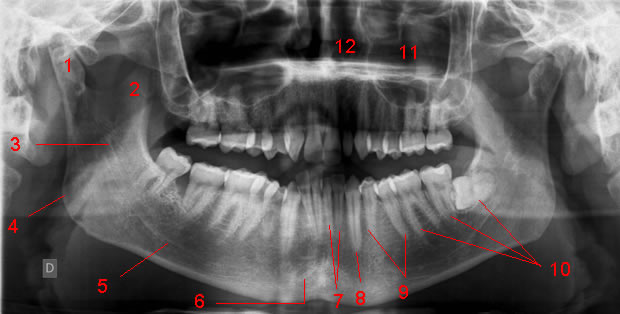
1, Condyle (Mandible.) 2, Coronoid Processus (Mandible). 3, Ramus. 4, Angle of the mandibule. 5, Body. 6,Symphysis mentis. 7, Incisor. 8, Canine. 9, Premolar. 10, Molar. 11, Maxillary Sinus. 12, Nasal cavity. 13, Hard palate.
There are 32 permanent teeth in the adult. Each quadrant has 2 incisors, 1 canine, 2 premolars, 3 molars. Teeth are numbred from «mesial» to «distal».
| Right | Left | |
| Maxillary teeth | 18 17 16 15 14 13 12 11 | 21 22 23 24 25 26 27 28 |
| Mandibular teeth | 48 47 46 45 44 43 42 41 | 31 32 33 34 35 36 37 38 |
No comments:
Post a Comment