- (See separate nerve articles for details on divisions proximal to the elbow and distal to the wrist; see Brachial plexus for the origins of the median, radial and ulnar nerves)
- Median nerve – principle nerve of the anterior compartment (PT, FCR, PL, FDS).
- anterior interosseous nerve (supplies FPL, lat. 1/2 of FDP, PQ).
- Radial nerve – supplies muscles of the posterior compartment (ECRL, ECRB).
- Superficial branch of radial nerve
- Deep branch of radial nerve, becomes Posterior interosseus nerve and supplies muscles of the posterior compartment (ED, EDM,ECU, APL, EPB, EPL, EI).
- Ulnar nerve - supplies some medial muscles (FCU, med. 1/2 of FDP).
Median nerve
From Wikipedia, the free encyclopedia
Nerve: median nerve 
Diagram from Gray's anatomy, depicting the peripheral nerves of the upper extremity, amongst others the median nerve Latin nervus medianus Gray's subject #210 938 Innervates Anterior compartment of the forearm(with two exceptions), Thenar eminence, Lumbricals, skin of the hand From Lateral cord and Medial cord MeSH Median+Nerve
The median nerve is a nerve in humans and other animals. It is in the upper limb. It is one of the five main nerves originating from the brachial plexus.The median nerve is formed from parts of the medial and lateral cords of the brachial plexus, and continues down the arm to enter the forearm with the brachial artery.It originates from the brachial plexus with roots from C6, C7, C8, & T1.The median nerve is the only nerve that passes through the carpal tunnel, where it may be compressed to cause carpal tunnel syndrome.
Contents
[hide]
[edit]Course
[edit]Course in the upper arm and cubital fossa
After receiving inputs from both the lateral and medial roots of the corresponding cords of thebrachial plexus, the median nerve courses with brachial artery on medial side of arm betweenbiceps brachii and brachialis. At first lateral to the artery, it then crosses anteriorly to run medial to the artery in the distal arm and into the cubital fossa.Inside the cubital fossa the median nerve passes medial to the brachial artery, in front of the point of insertion of the brachialis muscle and deep to the biceps.The median nerve gives off an articular branch in the upper arm as it passes the elbow joint.[edit]Course and branches in the forearm
The median nerve arises from the cubital fossa and passes between the two heads of pronator teres. It then travels between flexor digitorum superficialis and flexor digitorum profundus before emerging between flexor digitorum superficialis and flexor carpi radialis.The unbranched portion of the median nerve (which arises from the cubital fossa) innervates muscles of superficial and intermediate groups of the anterior compartment except flexor carpi ulnaris.The median nerve does give off two branches as it courses through the forearm:- The anterior interosseous branch courses with the anterior interosseous artery and innervates all the muscles of the anterior compartment of the forearm except the flexor carpi ulnaris and the medial (ulnar) half of flexor digitorum profundus. It ends with its innervation of pronator quadratus.
- The palmar cutaneous branch of the median nerve arises at the distal part of the forearm. It supplies sensory innervation to the lateral aspect of the skin of the palm (but not the digits).
[edit]Branches in the hand
The median nerve enters the hand through the carpal tunnel, deep to the flexor retinaculum along with the tendons of flexor digitorum superficialis, flexor digitorum profundus, and flexor pollicis longus.From there it sends off several branches:- 1. Recurrent branch to muscles of the thenar compartment (the recurrent branch is also called "the million dollar nerve")[1]
- 2. Digital cutaneous branches to common palmar digital branch and proper palmar digital branch of the median nerve which supply the:
- a) lateral (radial) three and a half digits on the palmar side
- b) index, middle and ring finger on dorsum of the hand
The median nerve supplies motor innervation to the first and second lumbrical muscles.[edit]Distribution
[edit]Arm
The median nerve has no voluntary motor or cutaneous function in the (upper) arm. It gives vascular branches to the wall of the brachial artery. These vascular branches carry sympathetic fibers.[edit]Forearm
It innervates all of the flexors in the forearm except flexor carpi ulnaris and that part of flexor digitorum profundus that supplies the medial two digits. The latter two muscles are supplied by the ulnar nerve (specifically the Muscular branches of ulnar nerve).The main portion of the median nerve supplies the following muscles:Superficial group:Intermediate group:The anterior interosseus branch of the median nerve supplies the following muscles:Deep group:- Flexor digitorum profundus (only the lateral half)
- Flexor pollicis longus
- Pronator quadratus
[edit]Hand
In the hand, the median nerve supplies motor innervation to the 1st and 2nd lumbrical muscles. It also supplies the muscles of the thenar eminence by a recurrent thenar branch. The rest of the intrinsic muscles of the hand are supplied by the ulnar nerve.The median nerve innervates the skin of the palmar side of the thumb, the index and middle finger, half the ring finger, and the nail bed of these fingers. The lateral part of the palm is supplied by the palmar cutaneous branch of the median nerve, which leaves the nerve proximal to the wrist creases. This palmar cutaneous branch travels in a separate fascial groove adjacent to the flexor carpi radialis and then superficial to the flexor retinaculum. It is therefore spared in carpal tunnel syndrome.The muscles of the hand supplied by the median nerve can be remembered using the mnemonic, "LOAF" for Lumbricals 1 & 2, Opponens pollicis, Abductor pollicis brevis and Flexor pollicis brevis. [2][edit]Injury
Injury of median nerve at different levels cause different syndromes.- Injury of this nerve at a level above the elbow results in loss of pronation and a reduction in flexion of the hand at the wrist.
- Entrapment at the level of the elbow or the proximal forearm could be due to the pronator teres syndrome.
- Injury to the anterior interosseous branch in the forearm causes the anterior interosseous syndrome.
- Injury by compression at the carpal tunnel causes carpal tunnel syndrome.
- Severing the median nerve causes median claw hand.
- In the hand, thenar muscles are paralyzed and will atrophy over time. Opposition and flexion of the thumb are lost. The thumb and index finger are arrested in adduction and hyperextension. This appearance of the hand is collectively referred as 'ape hand deformity'.[1]
[edit]Additional images
[edit]References
Anterior interosseous nerve
From Wikipedia, the free encyclopedia
Nerve: Anterior interosseous nerve 
Nerves of the left upper extremity. (Volar interosseus labeled at center right.) 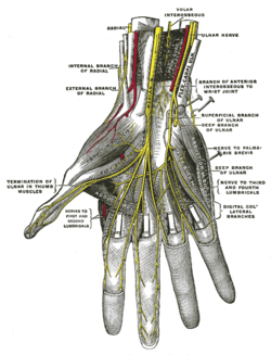
Deep palmar nerves. (Volar interosseous labeled at center top.) Latin nervus interosseus antebrachii anterior Gray's subject #210 938 From Median nerve
The anterior interosseous nerve (volar interosseous nerve) is a branch of the median nerve that supplies the deep muscles on the front of the forearm, except the ulnar half of theflexor digitorum profundus.It accompanies the anterior interosseous artery along the front of the interosseous membrane of the forearm, in the interval between the flexor pollicis longus and flexor digitorum profundus, supplying the whole of the former and the radial half of the latter, and ending below in thepronator quadratus and wrist joint.Many texts, for simplicity's sake, consider this nerve part of the median nerve.
Contents
[hide]
[edit]Innervation
The anterior interosseous nerve innervates 2.5 muscles:- flexor pollicis longus
- pronator quadratus
- the lateral half of flexor digitorum profundus that means lateral two out of the four tendons.
These muscles are in the deep level of the anterior compartment of the forearm.[edit]Injury
A branch of the median nerve, the anterior interosseous nerve (AIN) can be affected by either direct penetrating injury or compression in a fashion similar to carpal tunnel syndrome. The compression neuropathy is referred to an Anterior Interosseous Syndrome. As might be expected, the symptoms involve weakness in the muscle innervated by the AIN including the flexor digitorum profundus muscle to the index (and sometimes the middle) finger, the flexor pollicis longus muscle to the thumb and the pronator quadratus of the distal forearm. As opposed to carpal tunnel syndrome, the AIN has no sensory fibers and therefore no numbness associated with the Anterior Interosseous Syndrome. Non-surgical treatment consists of splinting, proximal tissue massage and anti-inflammatory drugs. Surgical treatment consists of releasing the compression on the nerve from surrounding structures.Pronator Syndrome is similar, but involves both the AIN as well as the median nerve proper.[edit]See also
[edit]External links
- Duke Orthopedics anterior_interosseous_branch_of_median_nerve
- anterior+interosseous+nerve at eMedicine Dictionary
- EatonHand ner-071
- -2006253539 at GPnotebook - "anterior interosseous nerve palsy"
- Image (see yellow arrow under "Findings")
This article was originally based on an entry from a public domain edition of Gray's Anatomy. As such, some of the information contained within it may be outdated.
[hide]
Nerves of upper limbs (primarily): the brachial plexus (C5-T1) (TA A14.2.03, GA 9.930) Supraclavicular Infraclavicular
median/lateral root: anterior interosseous · palmar · recurrent · common palmar digital (proper palmar digital) ulnar: muscular · palmar · dorsal (dorsal digital nerves) · superficial (common palmar digital, proper palmar digital) · deepmedian/medial root: see above
Other
Radial nerve
From Wikipedia, the free encyclopedia
Nerve: Radial nerve 
The suprascapular, axillary, and radial nerves. Latin nervus radialis Gray's subject #210 943 Innervates posterior compartment of the arm,posterior compartment of the forearm From posterior cord To posterior interosseous nerve MeSH Radial+Nerve
The radial nerve is a nerve in the human body that supplies the upper limb. It supplies thetriceps brachii muscle of the arm, as well as all 12 muscles in the posterior osteofascial compartment of the forearm, as well as the associated joints and overlying skin.It originates from the posterior cord of the brachial plexus with roots from C5, C6, C7, C8 & T1.The radial nerve and its branches supply the dorsal muscles, such as triceps brachii, the extrinsic extensors of the wrist and hands, and the cutaneous nerve supply to most of the back of the hand. (The ulnar nerve cutaneously innervates the back of the little finger and adjacent half of the ring finger).The radial nerve divides into a deep branch (which becomes the posterior interosseous nerve), and continues as the superficial branch which goes on to innervate the dorsum (back) of the hand.
Contents
[hide]
[edit]Course
The radial nerve originates as a terminal branch of the posterior cord of the brachial plexus. It goes through the arm, first in the posterior compartment of the arm, and later in the anterior compartment of the arm, and continues in the posterior compartment of the forearm.[edit]In arm
From the brachial plexus, it travels posteriorly through what is often called the triangular interval (US) or the triangular space of the axilla (UK).The radial nerve enters the arm behind the axillary artery/brachial artery, and it then travels posteriorly on the medial side of the arm.After giving off branches to the long and medial heads of the triceps brachii, it enters a groove on the humerus, the radial sulcus.Along with the deep brachial artery, the radial nerve winds around in the groove (between the medial and lateral heads of the triceps) towards the forearm, running laterally on the posterior aspect of the humerus.While in the groove, it gives off a branch to the lateral head of the triceps brachii.The radial nerve emerges from the groove on the lateral aspect of the humerus.At this point, it pierces the lateral intermuscular septum and enters the anterior compartment of the arm.It continues its journey inferiorly between the brachialis and brachioradialis muscles.When the radial nerve reaches the distal part of the humerus, it passes anterior to the lateral epicondyle and continues in the forearm.[edit]In forearm
In the forearm, it branches into a superficial branch (primarily sensory) and a deep branch (primarily motor).- The superficial branch of the radial nerve descends in the forearm under the brachioradialis. It eventually pierces the deep fascia near the back of the wrist.
- The deep branch of the radial nerve pierces the supinator muscle, after which it is known as the posterior interosseous nerve.
[edit]Branches/Innervations
The following are branches/innervations of the radial nerve (including the superficial branch of the radial nerve and the deep branch of the radial nerve/posterior interosseous nerve).[edit]Cutaneous
Cutaneous innervation by the radial nerve is provided by the following nerve branches:- Posterior cutaneous nerve of arm (originates in axilla)
- Inferior lateral cutaneous nerve of arm (originates in arm)
- Posterior cutaneous nerve of forearm (originates in arm)
The superficial branch of the radial nerve provides sensory innervation to much of the back of the hand, including the web of skin between the thumb and index finger.[edit]Motor
Posterior interosseous nerve (a continuation of the deep branch after the supinator):- Extensor digitorum
- Extensor digiti minimi
- Extensor carpi ulnaris
- Abductor pollicis longus
- Extensor pollicis brevis
- Extensor pollicis longus
- Extensor indicis
The radial nerve (and its deep branch) provides motor innervation to the muscles in the posterior compartment of the arm and forearm, which are mostly extensors. it also enters humerus[edit]Additional images
[edit]See also
- Muscular branches of the radial nerve
- Cutaneous branches of the radial nerve
- Superficial branch of the radial nerve
- Deep branch of the radial nerve
- Radial neuropathy
Vessels
Main article: Arterial tree of subclavian artery
Brachial artery
From Wikipedia, the free encyclopedia
Artery: Brachial artery The brachial artery. Right upper limb, anterior view, brachial arteryand elbow. Latin arteria brachialis Gray's subject #150 589 Supplies biceps brachii muscle, triceps brachii muscle Source axillary artery Branches Profunda brachii
Superior ulnar collateral artery
Inferior ulnar collateral artery Vein brachial vein MeSH Brachial+Artery
The brachial artery is the major blood vessel of the (upper) arm.It is the continuation of the axillary artery beyond the lower margin of teres major muscle. It continues down the ventral surface of the arm until it reaches the cubital fossa at the elbow. It then divides into the radial and ulnar arteries which run down the forearm. In some individuals, the bifurcation occurs much earlier and the ulnar and radial arteries extend through the upper arm. The pulse of the brachial artery is palpable on the anterior aspect of the elbow, medial to the tendon of the biceps, and, with the use of a stethoscope and sphygmomanometer (blood pressure cuff) often used to measure the blood pressure.The brachial artery is closely related to the median nerve; in proximal regions, the median nerve is immediately lateral to the brachial artery. Distally, the median nerve crosses the medial side of the brachial artery and lies anterior to the elbow joint.[edit]Branches
- Profunda brachii artery (deep brachial artery)
- Superior ulnar collateral artery
- Inferior ulnar collateral artery
- Radial artery (a terminal branch)
- Ulnar artery (a terminal branch)
- Nutrient branches to the humerus
as well as important anastomotic networks of the elbow and (as the axillary artery) the shoulder.The biceps head is lateral to the brachial artery. The median nerve is medial to the brachial artery for most of its course.[edit]Additional images
[edit]External links
- Dissection at mvm.ed.ac.uk
- Image at umich.edu - pulse
- Brachial+artery at eMedicine Dictionary
- lesson4arteriesofarm at The Anatomy Lesson by Wesley Norman (Georgetown University)
[hide]
List of arteries of upper limbs (TA A12.2.09, GA 6.575) Axillary
Shoulder Brachial
Before split forearm: ulnar recurrent (anterior, posterior) – common interosseous (anterior, posterior, recurrent)
wrist/carpus: dorsal carpal branch – palmar carpal branchhand: deep palmar branch Arches
Radial artery
From Wikipedia, the free encyclopedia
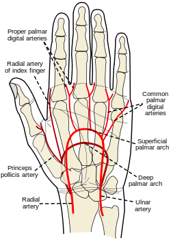
Artery: Radial artery Palm of left hand, showing position of skin creases and bones, and surface markings for the volar arches. Ulnar and radial arteries. Deep view. Latin A. Radialis Gray's subject #151 592 Source brachial artery Branches *in the forearm:
Radial recurrent artery
Palmar carpal branch of radial artery
Superficial palmar branch of the radial artery.
*At the wrist:
Dorsal carpal branch of radial artery
First dorsal metacarpal artery.
*In the hand:
Princeps pollicis artery
Radialis indicis
Deep palmar arch Vein radial vein MeSH Radial+Artery
In human anatomy, the radial artery is the main blood vessel, with oxygenated blood, of the lateral aspect of the forearm.
Contents
[hide]
[edit]Course
The radial artery arises from the bifurcation of the brachial artery in the cubital fossa. It runs distally on the anterior part of the forearm. There, it serves as a landmark for the division between the anterior and posterior compartments of the forearm, with the posterior compartment beginning just lateral to the artery. The artery winds laterally around the wrist, passing through the anatomical snuff box and between the heads of the first dorsal interosseous muscle. It passes anteriorly between the heads of the adductor pollicis, and becomes the deep palmar arch, which joins with the deep branch of the ulnar artery.Along its course, it is accompanied by a similarly named vein, the radial vein.[edit]Branches
The named branches of the radial artery may be divided into three groups, corresponding with the three regions in which the vessel is situated.[edit]In the Forearm
- Radial recurrent artery - arises just after the radial artery comes off the brachial artery. It travels superiorly to anastomose with the radial collateral artery around the elbow joint
- Palmar carpal branch of radial artery - a small vessel which arises near the lower border of the pronator quadratus
- Superficial palmar branch of the radial artery - arises from the radial artery, just where this vessel is about to wind around the lateral side of the wrist.
[edit]At the Wrist
- Dorsal carpal branch of radial artery - a small vessel which arises beneath the extensor tendons of the thumb
- First dorsal metacarpal artery - arises just before the radial artery passes between the two heads of the first dorsal interosseous muscle and divides almost immediately into two branches which supply the adjacent sides of the thumb and index finger; the lateral side of the thumb receives a branch directly from the radial artery.
[edit]In the Hand
- Princeps pollicis artery - arises from the radial artery just as it turns medially to the deep part of the hand.
- Radialis indicis - arises close to the princeps pollicis. The two arteries may arise from a common trunk, the first palmar metacarpal artery.
- Deep palmar arch - terminal part of radial artery.
[edit]Clinical significance
The artery's pulse is palpable in the anatomical snuff box and on the anterior aspect of the arm over the carpal bones (where it is commonly used to assess the heart rate and cardiac rhythm).The radial artery is used for coronary artery bypass grafting and is growing in popularity among cardiac surgeons.[1] Recently, it has been shown to have a superior peri-operative and post-operative course when compared to saphenous vein grafts.[2][edit]See also
[edit]Additional images
[edit]References
Radial recurrent artery
From Wikipedia, the free encyclopedia
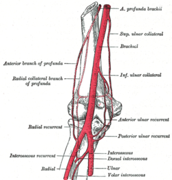
Artery: Radial recurrent artery Diagram of the anastomosis around the elbow-joint. (Radial recurrent labeled at center left.) The radial and ulnar arteries. (Radial recurrent labeled at center left.) Latin a. recurrens radialis Gray's subject #151 594 Source radial artery
The radial recurrent artery arises from the radial artery immediately below the elbow.It ascends between the branches of the radial nerve, lying on the Supinator and then between the Brachioradialis and Brachialis, supplying these muscles and the elbow-joint, and anastomosing with the terminal part of the profunda brachii.[edit]External links
- Radial+recurrent+artery at eMedicine Dictionary
- lesson4arteriesofarm at The Anatomy Lesson by Wesley Norman (Georgetown University)
- lesson4artofforearm at The Anatomy Lesson by Wesley Norman (Georgetown University)
This article was originally based on an entry from a public domain edition of Gray's Anatomy. As such, some of the information contained within it may be outdated.

This cardiovascular system article is a stub. You can help Wikipedia by expanding it.
[hid
Ulnar artery
From Wikipedia, the free encyclopedia
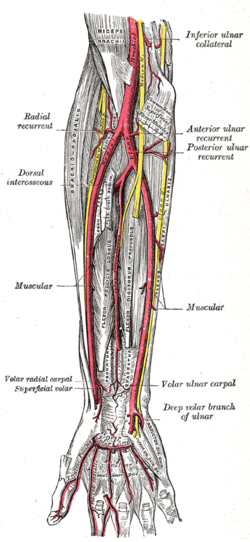
Artery: Ulnar artery Palm of left hand, showing position of skin creases and bones, and surface markings for the volar arches. Front of right upper extremity, showing surface markings for bones, arteries, and nerves. Latin A. Ulnaris Gray's subject #152 595 Source brachial artery Branches anterior ulnar recurrent artery
posterior ulnar recurrent artery
common interosseous artery(volar, dorsal)
muscular artery
volar carpal
dorsal carpal
deep volar
superficial volar arch Vein ulnar vein MeSH Ulnar+Artery
The ulnar artery is the main blood vessel, with oxygenated blood, of the medial aspect of theforearm. It arises from the brachial artery and terminates in the superficial palmar arch, which joins with the superficial branch of the radial artery. It is palpable on the anterior and medial aspect of the wrist.Along its course, it is accompanied by a similarly named vein or veins, the ulnar vein or ulnar veins.The ulnar artery, the larger of the two terminal branches of the brachial, begins a little below the bend of the elbow in the cubital fossa, and, passing obliquely downward, reaches the ulnar side of the forearm at a point about midway between the elbow and the wrist. It then runs along the ulnar border to the wrist, crosses the transverse carpal ligament on the radial side of the pisiform bone, and immediately beyond this bone divides into two branches, which enter into the formation of the superficial and deep volar arches.
Contents
[hide]
[edit]Relations
[edit]Forearm
In its upper half, it is deeply seated, being covered by the Pronator teres, Flexor carpi radialis,Palmaris longus, and Flexor digitorum sublimis; it lies upon the Brachialis and Flexor digitorum profundus.The median nerve is in relation with the medial side of the artery for about 2.5 cm. and then crosses the vessel, being separated from it by the ulnar head of the Pronator teres.In the lower half of the forearm it lies upon the Flexor digitorum profundus, being covered by the integument and the superficial and deep fasciæ, and placed between the Flexor carpi ulnaris and Flexor digitorum sublimis.It is accompanied by two venæ comitantes, and is overlapped in its middle third by the Flexor carpi ulnaris; the ulnar nerve lies on the medial side of the lower two-thirds of the artery, and the palmar cutaneous branch of the nerve descends on the lower part of the vessel to the palm of the hand.[edit]Wrist
At the wrist the ulnar artery is covered by the integument and the volar carpal ligament, and lies upon the transverse carpal ligament. On its medial side is the pisiform bone, and, somewhat behind the artery, the ulnar nerve.[edit]Peculiarities
The ulnar artery varies in its origin in the proportion of about one in thirteen cases; it may arise about 5 to 7 cm. below the elbow, but more frequently higher, the brachial being more often the source of origin than the axillary.Variations in the position of this vessel are more common than in the radial. When its origin is normal, the course of the vessel is rarely changed.When it arises high up, it is almost invariably superficial to the Flexor muscles in the forearm, lying commonly beneath the fascia, more rarely between the fascia and integument.In a few cases, its position was subcutaneous in the upper part of the forearm, and subaponeurotic in the lower part.[edit]See also
[edit]Additional images
[edit]External links
Pulmonary artery
From Wikipedia, the free encyclopedia
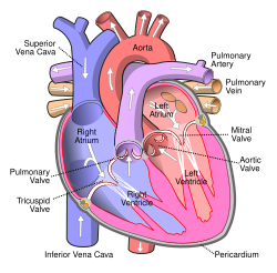
Artery: Pulmonary artery Anterior (frontal) view of the opened heart. White arrows indicate normal blood flow. (Pulmonary artery labeled at upper right.) Diagram of the alveoli with both cross-section and external view. Latin truncus pulmonalis, arteria pulmonalis Gray's subject #141 543 Source right ventricle Vein pulmonary vein Precursor truncus arteriosus MeSH Pulmonary+Artery
The pulmonary arteries carry blood from the heart to the lungs. They are the only arteries(other than umbilical arteries in the fetus) that carry deoxygenated blood.In the human heart, the pulmonary trunk (pulmonary artery or main pulmonary artery) begins at the base of the right ventricle. It is short and wide - approximately 5 cm (2 inches) in length and 3 cm (1.2 inches) in diameter. It then branches into two pulmonary arteries (left and right), which deliver de-oxygenated blood to the corresponding lung.
Contents
[hide]
[edit]Role in disease
Pulmonary hypertension occurs alone and as a consequence of a number of lung diseases. It can also be a consequence of heart disease (Eisenmenger's syndrome) but equally a cause (right-ventricular heart failure); it also occurs as a consequence of pulmonary embolism andscleroderma. It is characterised by reduced exercise tolerance. Severe forms, generally, have a dismal prognosis.[edit]Additional images
[edit]See also
Anterior ulnar recurrent artery
From Wikipedia, the free encyclopedia
Artery: Anterior ulnar recurrent artery Diagram of the anastomosis around the elbow-joint. (Anterior ulnar recurrent labeled at center right.) Ulnar and radial arteries. Deep view. (Anterior ulnar recurrent labeled at center right.) Latin ramus anterior arteriae recurrentis ulnaris Gray's subject #152 596 Source ulnar artery Branches inferior ulnar collateral artery
The anterior ulnar recurrent artery arises immediately below the elbow-joint, runs upward between the Brachialis and Pronator teres, supplies twigs to those muscles, and, in front of the medial epicondyle, anastomoses with the superior and inferior ulnar collateral arteries.[edit]External links
- lesson4arteriesofarm at The Anatomy Lesson by Wesley Norman (Georgetown University)
- lesson4artofforearm at The Anatomy Lesson by Wesley Norman (Georgetown University)
This article was originally based on an entry from a public domain edition of Gray's Anatomy. As such, some of the information contained within it may be outdated.

This cardiovascular system article is a stub. You can help Wikipedia by expanding it.
[hide]
Posterior ulnar recurrent artery
From Wikipedia, the free encyclopedia
Artery: Posterior ulnar recurrent artery Arteries of the back of the forearm and hand. (Posterior ulnar recurrent labeled at center left.) Ulnar and radial arteries. Deep view. (Posterior ulnar recurrent labeled at center right.) Latin ramus posterior arteriae recurrentis ulnaris Gray's subject #152 596 Source ulnar artery
The posterior ulnar recurrent artery is much larger than the anterior ulnar recurrent artery, and arises somewhat lower than it.It passes backward and medialward on the Flexor digitorum profundus, behind the Flexor digitorum sublimis, and ascends behind the medial epicondyle of the humerus.In the interval between this process and the olecranon, it lies beneath the Flexor carpi ulnaris, and ascending between the heads of that muscle, in relation with the ulnar nerve, it supplies the neighboring muscles and the elbow-joint, and anastomoses with the superior and inferior ulnar collateral and the interosseous recurrent arteries.[edit]External links
- lesson4arteriesofarm at The Anatomy Lesson by Wesley Norman (Georgetown University)
- lesson4artofforearm at The Anatomy Lesson by Wesley Norman (Georgetown University)
This article was originally based on an entry from a public domain edition of Gray's Anatomy. As such, some of the information contained within it may be outdated.

This cardiovascular system article is a stub. You can help Wikipedia by expanding it.
(See separate nerve articles for details on divisions proximal to the elbow and distal to the wrist; see Brachial plexus for the origins of the median, radial and ulnar nerves)
- Median nerve – principle nerve of the anterior compartment (PT, FCR, PL, FDS).
- anterior interosseous nerve (supplies FPL, lat. 1/2 of FDP, PQ).
- Radial nerve – supplies muscles of the posterior compartment (ECRL, ECRB).
- Superficial branch of radial nerve
- Deep branch of radial nerve, becomes Posterior interosseus nerve and supplies muscles of the posterior compartment (ED, EDM,ECU, APL, EPB, EPL, EI).
- Ulnar nerve - supplies some medial muscles (FCU, med. 1/2 of FDP).
Median nerve
From Wikipedia, the free encyclopedia
| Nerve: median nerve | |
|---|---|
 | |
| Diagram from Gray's anatomy, depicting the peripheral nerves of the upper extremity, amongst others the median nerve | |
| Latin | nervus medianus |
| Gray's | subject #210 938 |
| Innervates | Anterior compartment of the forearm(with two exceptions), Thenar eminence, Lumbricals, skin of the hand |
| From | Lateral cord and Medial cord |
| MeSH | Median+Nerve |
The median nerve is a nerve in humans and other animals. It is in the upper limb. It is one of the five main nerves originating from the brachial plexus.
The median nerve is formed from parts of the medial and lateral cords of the brachial plexus, and continues down the arm to enter the forearm with the brachial artery.
It originates from the brachial plexus with roots from C6, C7, C8, & T1.
The median nerve is the only nerve that passes through the carpal tunnel, where it may be compressed to cause carpal tunnel syndrome.
Contents[hide] |
[edit]Course
[edit]Course in the upper arm and cubital fossa
After receiving inputs from both the lateral and medial roots of the corresponding cords of thebrachial plexus, the median nerve courses with brachial artery on medial side of arm betweenbiceps brachii and brachialis. At first lateral to the artery, it then crosses anteriorly to run medial to the artery in the distal arm and into the cubital fossa.
Inside the cubital fossa the median nerve passes medial to the brachial artery, in front of the point of insertion of the brachialis muscle and deep to the biceps.
The median nerve gives off an articular branch in the upper arm as it passes the elbow joint.
[edit]Course and branches in the forearm
The median nerve arises from the cubital fossa and passes between the two heads of pronator teres. It then travels between flexor digitorum superficialis and flexor digitorum profundus before emerging between flexor digitorum superficialis and flexor carpi radialis.
The unbranched portion of the median nerve (which arises from the cubital fossa) innervates muscles of superficial and intermediate groups of the anterior compartment except flexor carpi ulnaris.
The median nerve does give off two branches as it courses through the forearm:
- The anterior interosseous branch courses with the anterior interosseous artery and innervates all the muscles of the anterior compartment of the forearm except the flexor carpi ulnaris and the medial (ulnar) half of flexor digitorum profundus. It ends with its innervation of pronator quadratus.
- The palmar cutaneous branch of the median nerve arises at the distal part of the forearm. It supplies sensory innervation to the lateral aspect of the skin of the palm (but not the digits).
[edit]Branches in the hand
The median nerve enters the hand through the carpal tunnel, deep to the flexor retinaculum along with the tendons of flexor digitorum superficialis, flexor digitorum profundus, and flexor pollicis longus.
From there it sends off several branches:
- 1. Recurrent branch to muscles of the thenar compartment (the recurrent branch is also called "the million dollar nerve")[1]
- 2. Digital cutaneous branches to common palmar digital branch and proper palmar digital branch of the median nerve which supply the:
- a) lateral (radial) three and a half digits on the palmar side
- b) index, middle and ring finger on dorsum of the hand
The median nerve supplies motor innervation to the first and second lumbrical muscles.
[edit]Distribution
[edit]Arm
The median nerve has no voluntary motor or cutaneous function in the (upper) arm. It gives vascular branches to the wall of the brachial artery. These vascular branches carry sympathetic fibers.
[edit]Forearm
It innervates all of the flexors in the forearm except flexor carpi ulnaris and that part of flexor digitorum profundus that supplies the medial two digits. The latter two muscles are supplied by the ulnar nerve (specifically the Muscular branches of ulnar nerve).
The main portion of the median nerve supplies the following muscles:
Superficial group:
Intermediate group:
The anterior interosseus branch of the median nerve supplies the following muscles:
Deep group:
- Flexor digitorum profundus (only the lateral half)
- Flexor pollicis longus
- Pronator quadratus
[edit]Hand
In the hand, the median nerve supplies motor innervation to the 1st and 2nd lumbrical muscles. It also supplies the muscles of the thenar eminence by a recurrent thenar branch. The rest of the intrinsic muscles of the hand are supplied by the ulnar nerve.
The median nerve innervates the skin of the palmar side of the thumb, the index and middle finger, half the ring finger, and the nail bed of these fingers. The lateral part of the palm is supplied by the palmar cutaneous branch of the median nerve, which leaves the nerve proximal to the wrist creases. This palmar cutaneous branch travels in a separate fascial groove adjacent to the flexor carpi radialis and then superficial to the flexor retinaculum. It is therefore spared in carpal tunnel syndrome.
The muscles of the hand supplied by the median nerve can be remembered using the mnemonic, "LOAF" for Lumbricals 1 & 2, Opponens pollicis, Abductor pollicis brevis and Flexor pollicis brevis. [2]
[edit]Injury
Injury of median nerve at different levels cause different syndromes.
- Injury of this nerve at a level above the elbow results in loss of pronation and a reduction in flexion of the hand at the wrist.
- Entrapment at the level of the elbow or the proximal forearm could be due to the pronator teres syndrome.
- Injury to the anterior interosseous branch in the forearm causes the anterior interosseous syndrome.
- Injury by compression at the carpal tunnel causes carpal tunnel syndrome.
- Severing the median nerve causes median claw hand.
- In the hand, thenar muscles are paralyzed and will atrophy over time. Opposition and flexion of the thumb are lost. The thumb and index finger are arrested in adduction and hyperextension. This appearance of the hand is collectively referred as 'ape hand deformity'.[1]
[edit]Additional images
[edit]References
Anterior interosseous nerve
From Wikipedia, the free encyclopedia
| Nerve: Anterior interosseous nerve | |
|---|---|
 | |
| Nerves of the left upper extremity. (Volar interosseus labeled at center right.) | |
 | |
| Deep palmar nerves. (Volar interosseous labeled at center top.) | |
| Latin | nervus interosseus antebrachii anterior |
| Gray's | subject #210 938 |
| From | Median nerve |
The anterior interosseous nerve (volar interosseous nerve) is a branch of the median nerve that supplies the deep muscles on the front of the forearm, except the ulnar half of theflexor digitorum profundus.
It accompanies the anterior interosseous artery along the front of the interosseous membrane of the forearm, in the interval between the flexor pollicis longus and flexor digitorum profundus, supplying the whole of the former and the radial half of the latter, and ending below in thepronator quadratus and wrist joint.
Many texts, for simplicity's sake, consider this nerve part of the median nerve.
Contents[hide] |
[edit]Innervation
The anterior interosseous nerve innervates 2.5 muscles:
- flexor pollicis longus
- pronator quadratus
- the lateral half of flexor digitorum profundus that means lateral two out of the four tendons.
These muscles are in the deep level of the anterior compartment of the forearm.
[edit]Injury
A branch of the median nerve, the anterior interosseous nerve (AIN) can be affected by either direct penetrating injury or compression in a fashion similar to carpal tunnel syndrome. The compression neuropathy is referred to an Anterior Interosseous Syndrome. As might be expected, the symptoms involve weakness in the muscle innervated by the AIN including the flexor digitorum profundus muscle to the index (and sometimes the middle) finger, the flexor pollicis longus muscle to the thumb and the pronator quadratus of the distal forearm. As opposed to carpal tunnel syndrome, the AIN has no sensory fibers and therefore no numbness associated with the Anterior Interosseous Syndrome. Non-surgical treatment consists of splinting, proximal tissue massage and anti-inflammatory drugs. Surgical treatment consists of releasing the compression on the nerve from surrounding structures.Pronator Syndrome is similar, but involves both the AIN as well as the median nerve proper.
[edit]See also
[edit]External links
- Duke Orthopedics anterior_interosseous_branch_of_median_nerve
- anterior+interosseous+nerve at eMedicine Dictionary
- EatonHand ner-071
- -2006253539 at GPnotebook - "anterior interosseous nerve palsy"
- Image (see yellow arrow under "Findings")
This article was originally based on an entry from a public domain edition of Gray's Anatomy. As such, some of the information contained within it may be outdated.
| ||||||||||||||||||||||||
From Wikipedia, the free encyclopedia
| Nerve: Radial nerve | |
|---|---|
 | |
| The suprascapular, axillary, and radial nerves. | |
| Latin | nervus radialis |
| Gray's | subject #210 943 |
| Innervates | posterior compartment of the arm,posterior compartment of the forearm |
| From | posterior cord |
| To | posterior interosseous nerve |
| MeSH | Radial+Nerve |
The radial nerve is a nerve in the human body that supplies the upper limb. It supplies thetriceps brachii muscle of the arm, as well as all 12 muscles in the posterior osteofascial compartment of the forearm, as well as the associated joints and overlying skin.
It originates from the posterior cord of the brachial plexus with roots from C5, C6, C7, C8 & T1.
The radial nerve and its branches supply the dorsal muscles, such as triceps brachii, the extrinsic extensors of the wrist and hands, and the cutaneous nerve supply to most of the back of the hand. (The ulnar nerve cutaneously innervates the back of the little finger and adjacent half of the ring finger).
The radial nerve divides into a deep branch (which becomes the posterior interosseous nerve), and continues as the superficial branch which goes on to innervate the dorsum (back) of the hand.
Contents[hide] |
[edit]Course
The radial nerve originates as a terminal branch of the posterior cord of the brachial plexus. It goes through the arm, first in the posterior compartment of the arm, and later in the anterior compartment of the arm, and continues in the posterior compartment of the forearm.
[edit]In arm
From the brachial plexus, it travels posteriorly through what is often called the triangular interval (US) or the triangular space of the axilla (UK).
The radial nerve enters the arm behind the axillary artery/brachial artery, and it then travels posteriorly on the medial side of the arm.
After giving off branches to the long and medial heads of the triceps brachii, it enters a groove on the humerus, the radial sulcus.
Along with the deep brachial artery, the radial nerve winds around in the groove (between the medial and lateral heads of the triceps) towards the forearm, running laterally on the posterior aspect of the humerus.
While in the groove, it gives off a branch to the lateral head of the triceps brachii.
The radial nerve emerges from the groove on the lateral aspect of the humerus.
At this point, it pierces the lateral intermuscular septum and enters the anterior compartment of the arm.
It continues its journey inferiorly between the brachialis and brachioradialis muscles.
When the radial nerve reaches the distal part of the humerus, it passes anterior to the lateral epicondyle and continues in the forearm.
[edit]In forearm
In the forearm, it branches into a superficial branch (primarily sensory) and a deep branch (primarily motor).
- The superficial branch of the radial nerve descends in the forearm under the brachioradialis. It eventually pierces the deep fascia near the back of the wrist.
- The deep branch of the radial nerve pierces the supinator muscle, after which it is known as the posterior interosseous nerve.
[edit]Branches/Innervations
The following are branches/innervations of the radial nerve (including the superficial branch of the radial nerve and the deep branch of the radial nerve/posterior interosseous nerve).
[edit]Cutaneous
Cutaneous innervation by the radial nerve is provided by the following nerve branches:
- Posterior cutaneous nerve of arm (originates in axilla)
- Inferior lateral cutaneous nerve of arm (originates in arm)
- Posterior cutaneous nerve of forearm (originates in arm)
The superficial branch of the radial nerve provides sensory innervation to much of the back of the hand, including the web of skin between the thumb and index finger.
[edit]Motor
Posterior interosseous nerve (a continuation of the deep branch after the supinator):
- Extensor digitorum
- Extensor digiti minimi
- Extensor carpi ulnaris
- Abductor pollicis longus
- Extensor pollicis brevis
- Extensor pollicis longus
- Extensor indicis
The radial nerve (and its deep branch) provides motor innervation to the muscles in the posterior compartment of the arm and forearm, which are mostly extensors. it also enters humerus
[edit]Additional images
[edit]See also
- Muscular branches of the radial nerve
- Cutaneous branches of the radial nerve
- Superficial branch of the radial nerve
- Deep branch of the radial nerve
- Radial neuropathy
Vessels
Main article: Arterial tree of subclavian artery
Brachial artery
From Wikipedia, the free encyclopedia
| Artery: Brachial artery | |
|---|---|
| The brachial artery. | |
| Right upper limb, anterior view, brachial arteryand elbow. | |
| Latin | arteria brachialis |
| Gray's | subject #150 589 |
| Supplies | biceps brachii muscle, triceps brachii muscle |
| Source | axillary artery |
| Branches | Profunda brachii Superior ulnar collateral artery Inferior ulnar collateral artery |
| Vein | brachial vein |
| MeSH | Brachial+Artery |
The brachial artery is the major blood vessel of the (upper) arm.
It is the continuation of the axillary artery beyond the lower margin of teres major muscle. It continues down the ventral surface of the arm until it reaches the cubital fossa at the elbow. It then divides into the radial and ulnar arteries which run down the forearm. In some individuals, the bifurcation occurs much earlier and the ulnar and radial arteries extend through the upper arm. The pulse of the brachial artery is palpable on the anterior aspect of the elbow, medial to the tendon of the biceps, and, with the use of a stethoscope and sphygmomanometer (blood pressure cuff) often used to measure the blood pressure.
The brachial artery is closely related to the median nerve; in proximal regions, the median nerve is immediately lateral to the brachial artery. Distally, the median nerve crosses the medial side of the brachial artery and lies anterior to the elbow joint.
[edit]Branches
- Profunda brachii artery (deep brachial artery)
- Superior ulnar collateral artery
- Inferior ulnar collateral artery
- Radial artery (a terminal branch)
- Ulnar artery (a terminal branch)
- Nutrient branches to the humerus
as well as important anastomotic networks of the elbow and (as the axillary artery) the shoulder.
The biceps head is lateral to the brachial artery. The median nerve is medial to the brachial artery for most of its course.
[edit]Additional images
[edit]External links
- Dissection at mvm.ed.ac.uk
- Image at umich.edu - pulse
- Brachial+artery at eMedicine Dictionary
- lesson4arteriesofarm at The Anatomy Lesson by Wesley Norman (Georgetown University)
| ||||||||||||||||||||||||||
Radial artery
From Wikipedia, the free encyclopedia
| Artery: Radial artery | |
|---|---|
| Palm of left hand, showing position of skin creases and bones, and surface markings for the volar arches. | |
| Ulnar and radial arteries. Deep view. | |
| Latin | A. Radialis |
| Gray's | subject #151 592 |
| Source | brachial artery |
| Branches | *in the forearm: Radial recurrent artery Palmar carpal branch of radial artery Superficial palmar branch of the radial artery. *At the wrist: Dorsal carpal branch of radial artery First dorsal metacarpal artery. *In the hand: Princeps pollicis artery Radialis indicis Deep palmar arch |
| Vein | radial vein |
| MeSH | Radial+Artery |
In human anatomy, the radial artery is the main blood vessel, with oxygenated blood, of the lateral aspect of the forearm.
Contents[hide] |
[edit]Course
The radial artery arises from the bifurcation of the brachial artery in the cubital fossa. It runs distally on the anterior part of the forearm. There, it serves as a landmark for the division between the anterior and posterior compartments of the forearm, with the posterior compartment beginning just lateral to the artery. The artery winds laterally around the wrist, passing through the anatomical snuff box and between the heads of the first dorsal interosseous muscle. It passes anteriorly between the heads of the adductor pollicis, and becomes the deep palmar arch, which joins with the deep branch of the ulnar artery.
Along its course, it is accompanied by a similarly named vein, the radial vein.
[edit]Branches
The named branches of the radial artery may be divided into three groups, corresponding with the three regions in which the vessel is situated.
[edit]In the Forearm
- Radial recurrent artery - arises just after the radial artery comes off the brachial artery. It travels superiorly to anastomose with the radial collateral artery around the elbow joint
- Palmar carpal branch of radial artery - a small vessel which arises near the lower border of the pronator quadratus
- Superficial palmar branch of the radial artery - arises from the radial artery, just where this vessel is about to wind around the lateral side of the wrist.
[edit]At the Wrist
- Dorsal carpal branch of radial artery - a small vessel which arises beneath the extensor tendons of the thumb
- First dorsal metacarpal artery - arises just before the radial artery passes between the two heads of the first dorsal interosseous muscle and divides almost immediately into two branches which supply the adjacent sides of the thumb and index finger; the lateral side of the thumb receives a branch directly from the radial artery.
[edit]In the Hand
- Princeps pollicis artery - arises from the radial artery just as it turns medially to the deep part of the hand.
- Radialis indicis - arises close to the princeps pollicis. The two arteries may arise from a common trunk, the first palmar metacarpal artery.
- Deep palmar arch - terminal part of radial artery.
[edit]Clinical significance
The artery's pulse is palpable in the anatomical snuff box and on the anterior aspect of the arm over the carpal bones (where it is commonly used to assess the heart rate and cardiac rhythm).
The radial artery is used for coronary artery bypass grafting and is growing in popularity among cardiac surgeons.[1] Recently, it has been shown to have a superior peri-operative and post-operative course when compared to saphenous vein grafts.[2]
[edit]See also
[edit]Additional images
[edit]References
Radial recurrent artery
From Wikipedia, the free encyclopedia
| Artery: Radial recurrent artery | |
|---|---|
| Diagram of the anastomosis around the elbow-joint. (Radial recurrent labeled at center left.) | |
| The radial and ulnar arteries. (Radial recurrent labeled at center left.) | |
| Latin | a. recurrens radialis |
| Gray's | subject #151 594 |
| Source | radial artery |
The radial recurrent artery arises from the radial artery immediately below the elbow.
It ascends between the branches of the radial nerve, lying on the Supinator and then between the Brachioradialis and Brachialis, supplying these muscles and the elbow-joint, and anastomosing with the terminal part of the profunda brachii.
[edit]External links
- Radial+recurrent+artery at eMedicine Dictionary
- lesson4arteriesofarm at The Anatomy Lesson by Wesley Norman (Georgetown University)
- lesson4artofforearm at The Anatomy Lesson by Wesley Norman (Georgetown University)
This article was originally based on an entry from a public domain edition of Gray's Anatomy. As such, some of the information contained within it may be outdated.
| This cardiovascular system article is a stub. You can help Wikipedia by expanding it. |
| ||
Ulnar artery
From Wikipedia, the free encyclopedia
| Artery: Ulnar artery | |
|---|---|
| Palm of left hand, showing position of skin creases and bones, and surface markings for the volar arches. | |
| Front of right upper extremity, showing surface markings for bones, arteries, and nerves. | |
| Latin | A. Ulnaris |
| Gray's | subject #152 595 |
| Source | brachial artery |
| Branches | anterior ulnar recurrent artery posterior ulnar recurrent artery common interosseous artery(volar, dorsal) muscular artery volar carpal dorsal carpal deep volar superficial volar arch |
| Vein | ulnar vein |
| MeSH | Ulnar+Artery |
The ulnar artery is the main blood vessel, with oxygenated blood, of the medial aspect of theforearm. It arises from the brachial artery and terminates in the superficial palmar arch, which joins with the superficial branch of the radial artery. It is palpable on the anterior and medial aspect of the wrist.
Along its course, it is accompanied by a similarly named vein or veins, the ulnar vein or ulnar veins.
The ulnar artery, the larger of the two terminal branches of the brachial, begins a little below the bend of the elbow in the cubital fossa, and, passing obliquely downward, reaches the ulnar side of the forearm at a point about midway between the elbow and the wrist. It then runs along the ulnar border to the wrist, crosses the transverse carpal ligament on the radial side of the pisiform bone, and immediately beyond this bone divides into two branches, which enter into the formation of the superficial and deep volar arches.
Contents[hide] |
[edit]Relations
[edit]Forearm
In its upper half, it is deeply seated, being covered by the Pronator teres, Flexor carpi radialis,Palmaris longus, and Flexor digitorum sublimis; it lies upon the Brachialis and Flexor digitorum profundus.
The median nerve is in relation with the medial side of the artery for about 2.5 cm. and then crosses the vessel, being separated from it by the ulnar head of the Pronator teres.
In the lower half of the forearm it lies upon the Flexor digitorum profundus, being covered by the integument and the superficial and deep fasciæ, and placed between the Flexor carpi ulnaris and Flexor digitorum sublimis.
It is accompanied by two venæ comitantes, and is overlapped in its middle third by the Flexor carpi ulnaris; the ulnar nerve lies on the medial side of the lower two-thirds of the artery, and the palmar cutaneous branch of the nerve descends on the lower part of the vessel to the palm of the hand.
[edit]Wrist
At the wrist the ulnar artery is covered by the integument and the volar carpal ligament, and lies upon the transverse carpal ligament. On its medial side is the pisiform bone, and, somewhat behind the artery, the ulnar nerve.
[edit]Peculiarities
The ulnar artery varies in its origin in the proportion of about one in thirteen cases; it may arise about 5 to 7 cm. below the elbow, but more frequently higher, the brachial being more often the source of origin than the axillary.
Variations in the position of this vessel are more common than in the radial. When its origin is normal, the course of the vessel is rarely changed.
When it arises high up, it is almost invariably superficial to the Flexor muscles in the forearm, lying commonly beneath the fascia, more rarely between the fascia and integument.
In a few cases, its position was subcutaneous in the upper part of the forearm, and subaponeurotic in the lower part.
[edit]See also
[edit]Additional images
[edit]External links
Pulmonary artery
From Wikipedia, the free encyclopedia
| Artery: Pulmonary artery | |
|---|---|
| Anterior (frontal) view of the opened heart. White arrows indicate normal blood flow. (Pulmonary artery labeled at upper right.) | |
| Diagram of the alveoli with both cross-section and external view. | |
| Latin | truncus pulmonalis, arteria pulmonalis |
| Gray's | subject #141 543 |
| Source | right ventricle |
| Vein | pulmonary vein |
| Precursor | truncus arteriosus |
| MeSH | Pulmonary+Artery |
The pulmonary arteries carry blood from the heart to the lungs. They are the only arteries(other than umbilical arteries in the fetus) that carry deoxygenated blood.
In the human heart, the pulmonary trunk (pulmonary artery or main pulmonary artery) begins at the base of the right ventricle. It is short and wide - approximately 5 cm (2 inches) in length and 3 cm (1.2 inches) in diameter. It then branches into two pulmonary arteries (left and right), which deliver de-oxygenated blood to the corresponding lung.
Contents[hide] |
[edit]Role in disease
Pulmonary hypertension occurs alone and as a consequence of a number of lung diseases. It can also be a consequence of heart disease (Eisenmenger's syndrome) but equally a cause (right-ventricular heart failure); it also occurs as a consequence of pulmonary embolism andscleroderma. It is characterised by reduced exercise tolerance. Severe forms, generally, have a dismal prognosis.
[edit]Additional images
[edit]See also
Anterior ulnar recurrent artery
From Wikipedia, the free encyclopedia
| Artery: Anterior ulnar recurrent artery | |
|---|---|
| Diagram of the anastomosis around the elbow-joint. (Anterior ulnar recurrent labeled at center right.) | |
| Ulnar and radial arteries. Deep view. (Anterior ulnar recurrent labeled at center right.) | |
| Latin | ramus anterior arteriae recurrentis ulnaris |
| Gray's | subject #152 596 |
| Source | ulnar artery |
| Branches | inferior ulnar collateral artery |
The anterior ulnar recurrent artery arises immediately below the elbow-joint, runs upward between the Brachialis and Pronator teres, supplies twigs to those muscles, and, in front of the medial epicondyle, anastomoses with the superior and inferior ulnar collateral arteries.
[edit]External links
- lesson4arteriesofarm at The Anatomy Lesson by Wesley Norman (Georgetown University)
- lesson4artofforearm at The Anatomy Lesson by Wesley Norman (Georgetown University)
This article was originally based on an entry from a public domain edition of Gray's Anatomy. As such, some of the information contained within it may be outdated.
| This cardiovascular system article is a stub. You can help Wikipedia by expanding it. |
| ||
Posterior ulnar recurrent artery
From Wikipedia, the free encyclopedia
| Artery: Posterior ulnar recurrent artery | |
|---|---|
| Arteries of the back of the forearm and hand. (Posterior ulnar recurrent labeled at center left.) | |
| Ulnar and radial arteries. Deep view. (Posterior ulnar recurrent labeled at center right.) | |
| Latin | ramus posterior arteriae recurrentis ulnaris |
| Gray's | subject #152 596 |
| Source | ulnar artery |
The posterior ulnar recurrent artery is much larger than the anterior ulnar recurrent artery, and arises somewhat lower than it.
It passes backward and medialward on the Flexor digitorum profundus, behind the Flexor digitorum sublimis, and ascends behind the medial epicondyle of the humerus.
In the interval between this process and the olecranon, it lies beneath the Flexor carpi ulnaris, and ascending between the heads of that muscle, in relation with the ulnar nerve, it supplies the neighboring muscles and the elbow-joint, and anastomoses with the superior and inferior ulnar collateral and the interosseous recurrent arteries.
[edit]External links
- lesson4arteriesofarm at The Anatomy Lesson by Wesley Norman (Georgetown University)
- lesson4artofforearm at The Anatomy Lesson by Wesley Norman (Georgetown University)
This article was originally based on an entry from a public domain edition of Gray's Anatomy. As such, some of the information contained within it may be outdated.
| This cardiovascular system article is a stub. You can help Wikipedia by expanding it. |













No comments:
Post a Comment