Showing posts with label CARDIAC RADIOLOGY. Show all posts

Friday, August 13, 2010

Recognizing A Technically Adequate Chest X-ray


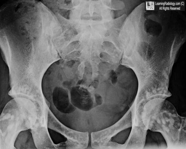
What is the most likely diagnosis?

  • 37 year-old female with abdominal pain


Frontal radiograph of the pelvis
  1. Osteoblastic breast metastases
  2. Dermoid of the ovary
  3. Osteopoikilosis
  4. Pelvic fracture
  5. Multiple myeloma
Additional Images - Axial CT scan of the pelvis


Recognizing
A Technically
Adequate Chest X-ray

Factors to Evaluate
lPenetration
lInspiration
lRotation
lAngulation


You should be able to
just see the thoracic
spine through the
heart.


Wednesday, August 11, 2010

Bronchiolitis

Bronchiolitis


Bronchiolitis
Classification and external resources

chest X-ray demonstrating lung hyperinflation with a flattened diaphragm and bilateralatelectasis in the right apical and left basal regions in a 16-day-old infant with severe bronchiolitis.
ICD-10J21.
ICD-9466.1
DiseasesDB1701
MedlinePlus000975
eMedicineemerg/365
MeSHD001988
Bronchiolitis is inflammation of the bronchioles, the smallest air passages of the lungs. This inflammation is usually caused by viruses.

Contents

 [hide]

[edit]Causes

The term usually refers to acute viral bronchiolitis, a common disease in infancy. This is most commonly caused by respiratory syncytial virus (RSV, also known as human pneumovirus). (J21.0)
Other viruses which may cause this illness include metapneumovirusinfluenzaparainfluenza,coronavirusadenovirus, and rhinovirus.
The American Academy of Pediatrics has published a clinical practice guideline for theDiagnosis and Management of Bronchiolitis, including a review of the evidence and recommendations.

[edit]Diagnosis and Recovery

In a typical case, an infant under two years of age develops cough, wheeze, and shortness of breath over one or two days. The diagnosis is made by clinical examination. Chest X-ray is sometimes useful to exclude pneumonia, but not indicated in routine cases.
Testing for specific viral cause (e.g. RSV by nasopharyngeal aspirate) can be done but has little effect on management. In a systematic review, RSV testing by direct immunofluorescence testing on nasopharyngeal aspirate had a sensitivity of 61% and specificity of 89%.[1]Identification of RSV-positive patients can be helpful for:
  • disease surveillance
  • grouping ("cohorting") patients together in hospital wards as to prevent cross infection
  • predicting whether the disease course has peaked yet
  • reducing the need for other diagnostic procedures (by providing confidence that a cause has been identified).
The infant may be breathless for several days. After the acute illness, it is common for the airways to remain sensitive for several weeks, leading to recurrent cough and wheeze.
There is a possible link with later asthma: possible explanations are that bronchiolitis causes asthma by inducing long term inflammation, or that children who are destined to be asthmatic are more susceptible to develop bronchiolitis.
Bronchiolitis Obliterans occurs rarely in rheumatoid lung disease when small airway obstruction develops into a necrotizing bronchiolitis.

[edit]Treatment

There is no effective specific treatment for bronchiolitis. Therapy is principally supportive.[2]

[edit]Conservative measures

Frequent small feeds are encouraged to maintain hydration as evidenced by good urine output, and sometimes oxygen may be required to maintain blood oxygen levels. Suction of the nasopharynx is often performed to maintain a clear airway. In severe cases the infant may need to be fed via a nasogastric tube or it may even need intravenous fluids. In extreme cases, mechanical ventilation (for example, using CPAP) might be necessary.

[edit]Bronchodilators

Bronchodilator drugs such as salbutamol/albuterol or ipratropium are no longer recommended, but many clinicians offer a trial dose to see if there is any benefit (especially if there is a family history of asthma, since it can be difficult to clinically distinguish bronchiolitis from a viral-induced asthma). Racemic epinephrine is another drug that is sometimes given.

[edit]Hypertonic saline

There is some interest in the use of hypertonic saline in bronchiolitis. Initially recommended for use in cystic fibrosis patients, it is speculated to increase hydration of secretions, thus facilitating their removal. [3] In a study of emergency room patients with bronchiolitis, a comparison of nebulized 3% saline/epinephrine with 0.9% saline/epinephrine showed no difference in effectiveness.[4] However, in another study looking at the inpatient population, aerosolized 3% saline soln/1.5mg epinephrine was shown to reduce the length of hospital stay among non-asthmatic, non-severely ill infants with viral bronchiolitis as compared to aerosolized 0.9% saline soln/1.5mg epinephrine according to a randomized controlled trial. Length of stay for aerosolized 3% saline soln/1.5mg epinephrine was 3 +/- 1.2 days, which was significantly shorter (by 25%) than length of stay for patients treated with 0.9% saline soln/1.5mg epinephrine which was 4 +/- 1.9 days (p < 0.05).[5]

[edit]Non effective

Ribavirin is an antiviral drug which has a controversial role in treating RSV infection. There is no proven benefit but it is used sometimes for infants with pre-existing lung, heart or immune disease. Antibiotics are often given in case of a bacterial infection complicating bronchiolitis, but have no effect on the underlying viral infection.
Corticosteroids have no proven benefit in bronchiolitis treatment and are not advised.
DNAse has not been found to be effective.[6]

[edit]Complications

Middle ear bacterial infection
Development of asthma later (bronchial hyperactivity)

[edit]Prevention

In general, prevention of bronchiolitis relies on measures to reduce the spread of the viruses that cause respiratory infections (that is, handwashing, and avoiding exposure to those symptomatic with respiratory infections).
Premature infants, and others with certain major cardiac and respiratory disorders, can receive passive immunization with Palivizumab (amonoclonal antibody against RSV). This form of passive immunization therapy requires monthly injections every winter. Whether it could benefit infants with lung problems secondary to muscular dystrophies and other vulnerable groups is currently unknown


Introduction

Background

Bronchiolitis is an acute infectious disease of the lower respiratory tract that occurs primarily in young infants, most often in those aged 2-24 months.

Pathophysiology

Bronchiolitis is usually due to a viral infection of the small airways (bronchioles). Infection of bronchiolar respiratory and ciliated epithelial cells produces increased mucus secretion, cell death, and sloughing, followed by a peribronchiolar lymphocytic infiltrate and submucosal edema. The combination of debris and edema produces critical narrowing and obstruction of small airways.
Decreased ventilation of portions of the lung causes ventilation/perfusion mismatching, resulting in hypoxia. During the expiratory phase of respiration, further dynamic narrowing of the airways produces disproportionate airflow decrease and resultant air trapping. Work of breathing is increased due to increased end-expiratory lung volume and decreased lung compliance. Recovery of pulmonary epithelial cells occurs after 3-4 days, but cilia do not regenerate for about 2 weeks. The debris is cleared by macrophages.
Infection is spread by direct contact with respiratory secretions. In the United States, epidemics last 2-4 months beginning in November and peaking in January or February. While 93% of cases occur between November and early April, sporadic cases may occur throughout the year. Attack rates within families are as high as 45% and are higher in daycare centers. Rates of hospital-acquired infection range from 20-47%. Previous infection with the common etiologic viruses does not confer immunity. Reinfection is common.

Frequency

United States

Annual incidence is 11.4% in children younger than 1 year and 6% in those aged 1-2 years. The illness accounts for 4500 deaths and 90,000 hospital admissions per year. Prevalence may be higher in urban areas.
In children aged 2 years, approximately 95% have serologic evidence of past infection with the predominant causative agent, respiratory syncytial virus (RSV). Unfortunately, presence of antibodies to RSV does not confer immunity.

International

RSV is an important respiratory pathogen worldwide. The frequency of bronchiolitis in developed countries appears to be similar to that in the United States. Epidemiologic data for underdeveloped countries are incomplete. Peak incidence of bronchiolitis usually occurs during winter months in temperate climates and during the rainy season in tropical climates. In the United States, the peak occurs in February. Morbidity and mortality may be higher in less developed countries because of poor nutrition and lack of resources for supportive medical care.

Mortality/Morbidity

Significant morbidity is unusual.
  • Hospitalization is required in up to 2% of cases; most of those patients are younger than 6 months. These patients account for as many as 17% of all infant hospitalizations. Hospitalization is significantly more likely at altitudes above 2500 meters (8000 ft). Mechanical ventilation is required for 3-7% of admitted patients.
  • The mortality rate is 1-2% of all hospitalized patients and 3-4% for patients with underlying cardiac or pulmonary disease.
  • Most deaths occur in infants younger than 6 months.

Race

  • Race and socioeconomic status may affect the frequency of contracting bronchiolitis.
  • Lower socioeconomic status may increase the likelihood of hospitalization. Hospitalization rates are higher in Native American, Alaskan, and Hispanic populations, but it is not clear if this is due to more severe infection or a lower threshold for admission.

Sex

Bronchiolitis occurs as many as 1.25 times more frequently in males than in females.

Age

  • Although infection with etiologic agents may occur at any age, the clinical entity of bronchiolitis includes only infants and young children. Seventy-five percent of cases of bronchiolitis occur in children younger than 1 year, and 95% in children younger than 2 years. Incidence peaks in those aged 2-8 months.

Clinical

History

  • History and physical examination form the primary basis for the diagnosis of bronchiolitis.
  • Early symptoms are those of a viral upper respiratory tract infection (URI), including mild rhinorrhea, cough, and sometimes low-grade fever.
  • Adults, older children, and many infants do not progress beyond this stage of URI.
  • For the 40% of infants and young children who progress to lower respiratory tract involvement, paroxysmal cough and dyspnea develop within 1-2 days.
  • Other common symptoms include the following:
    • Fever
    • Increased work of breathing
    • Wheezing
    • Cyanosis
    • Grunting
    • Noisy breathing
    • Vomiting, especially post-tussive
    • Irritability
    • Poor feeding or anorexia

Physical

Most patients with bronchiolitis have the following signs:
  • Tachypnea, often at rates over 50-60 breaths per minute (most common physical sign)
  • Tachycardia
  • Fever, usually in the range of 38.5-39°C
  • Mild conjunctivitis or pharyngitis
  • Diffuse expiratory wheezing
  • Nasal flaring
  • Intercostal retractions
  • Cyanosis
  • Inspiratory crackles
  • Otitis media
  • Apnea, especially in infants younger than 6 weeks
  • Palpable liver and spleen from hyperinflation of the lungs and consequent depression of the diaphragm

Causes

RSV is the most common pathogen (85%), but other organisms occasionally produce a similar clinical picture.
  • Adenovirus (11%) occasionally causes a similar syndrome with a more virulent course.
  • Epidemics of bronchiolitis due to parainfluenza virus usually begin earlier in the year and tend to occur every other year.
  • Other less common etiologic agents include the following:
  • Ear, nose, or mouth inoculation
    • Exposure to an adult with a URI
    • Daycare exposure (significant)
    • Idiopathic


Wednesday, July 21, 2010

Osteoporosis Can Strike at Thirty


It is generally thought that osteoporosis occurs as a disease in older people. However, it can in actual fact strike at any age.
There is a risk for millions of people. The disease is associated with women, but as a matter of fact, men also suffer from the condition.   It true however, that women are four times more likely to develop osteoporosis than men.
Osteoporosis is a disease whereby the bones become fragile and brittle, which leads to a high risk of breakages or cracking of the bones.
Osteoporosis can affect any bone. However the most common areas are the hip bones, spine, ribs, pelvis, upper arms and wrists. 
There are generally no warning signs, symptoms or pain to indicate that an individual has osteoporosis, until the fracture occurs. These fractures can occur with a simple bump, strain or minor fall. It is for this reason that osteoporosis is very often referred to as the “silent disease”.
Changes in posture can be developed due to osteoporosis, such as a stoop in the back, weakness of the muscles, loss of height and bone deformity of the spine. Such fractures can lead to severe pain, disability, loss of independence and in serious cases, premature death.
An osteoporosis fracture is something that will be suffered by one in every two Caucasian women, and one in every eight men over the age of fifty years at some time in their remaining lifetime.
For women, in the first 5 to 7 years after menopause, it is possible to lose up to 20% of bone mass, which increases their chance of developing the disease.
The occurrence of osteoporosis happens when bones lose vital minerals, such as calcium, quicker than they can be replaced by the body, which leads to the loss of bone thickness.   Due to this, the bones become thinner and lose their density. Therefore fractures can occur when having the slightest bump or accident.
Some of the other factors also include:

  • Having a thin or small frame,

  • Anorexia nervosa,

  • Advanced age,

  • Post menopause, including early or induced menopause,

  • A family history of the disease,

  • Low testosterone in men,

  • Sedentary lifestyle,

  • Smoking,

  • The excessive consumption of alcohol.
Prevention
Osteoporosis is something which you are never to young to prepare yourself for.
The best foundation for prevention in later years is by building strong bones from early childhood and the teen years.                                 
By the age of twenty, an average woman will have acquired 98% of her skeletal mass.
There are three fundamental steps that a person should begin to take at any age, in order to minimize the chances of developing osteoporosis.
The first step, and the most important, is to increase the intake of calcium. It is necessary for most individuals to have an intake of 1,000 to 1,300 mgs of calcium on a daily basis in order to build and maintain healthy bones. There are many different sources of calcium, including calcium supplements. The intake of calcium should also be combined with Vitamin D. This assists the body in absorbing and retaining calcium. Again, there are many different sources of this valuable nutrient available.
Second, it is essential to lead a healthy lifestyle, with no heavy alcohol consumption and no smoking.                 
And third, regular exercise should be undertaken. Studies show that physical activity can greatly help to prevent osteoporosis. The body will benefit from exercise such as jogging, hiking, swimming, racquet sports and even weight lifting. However, for those individuals that are advancing in years, it is advised to seek medical advice on the size of weights and intensity of the exercise routines, as there is a danger of over doing it, which may result in fractures, if the disease is already present in the body.
According to medical experts, bone mass can be increased by as much as 20% in women by the age of thirty, if following a balanced diet and regular exercise routines.
Measurements of bone density can be taken in various parts of the body without any pain, discomfort or invasive procedure. These tests will establish an individuals’ rate of bone density loss and monitor the effects on a regular basis, as well as detecting the disease before experiencing a related fracture.
If an individual assumes that they have osteoporosis, then it is advised to consult a medical advisor and request a bone density test.


Thursday, July 8, 2010

An Approach to Arthritis


Definition
lDisease that affects bones on both
sides of the joint space and
lNarrows the space in between them


Arthritis or Not

Classification
lHypertrophic
nHallmarks
lBone production
lSclerosis
lInfectious
nHallmark
lDestruction of articular cortex
lErosive
nHallmark
lErosions
DJD
AVN
Hypertrophic Arthritis
l Degenerative arthritis
nPrimary
nSecondary
l Charcot arthropathy
1ยบ Degenerative Arthritis
lIntrinsic degeneration of articular
cartilage
lExcessive wear and tear
nMost commonly hips and knees
nLess commonly shoulders and elbows
1ยบ Degenerative Arthritis
lX-ray Findings
nNarrowing of  joint space
nSubchondral sclerosis
nMarginal osteophyte formation
nSubchondral cysts

1ยบ DJD of knees affects medial,
weight-bearing surface
1ยบ DJD of hips affects superior,
weight-bearing surface
1ยบ Degenerative Arthritis
Hands
lNot due to mechanical stress
lF:M  10:1
lMost often involves DIP joints
nSclerosis
nMarginal osteophyte formation
l1st MCP joint of thumb


1ยบ DJD of Hands


2ยบ Degenerative Arthritis
lAnother process destroys articular
cartilage
lDegenerative changes supervene
lHow to recognize
nAtypical locations (CPPD and knee)
nAtypical appearance (Marked DJD of 1 hip)
nAtypical age (DJD in 20 year-old
2ยบ Degenerative Arthritis
Causes
lTrauma
lInfection
lAvascular necrosis
lCPPD
lRA
lHemophilia


2ยบ Degenerative Arthritis
More Causes

lHemochromatosis
lAcromegaly
lOchronosis
lWilson's Disease
lBottom line: Any arthritis can end as
DJD


2ยบ DJD of right ankle following fracture
Calcium Pyrophosphate
Deposition Disease (CPPD)
nMay be idiopathic or associated with
lHyperparathyroidism, hemochromatosis
nSymmetric involvement: knees (most
common), wrists, MCPs
nSudden onset of pain and fever
nClinically
lTender, swollen, red, LOM
CPPD
Findings
nCalcification of articular cartilage
lKnee, hip, shoulder
lTriangular fibrocartilage of ulna
lSymphysis
nLarge subchondral cysts
nPreferential involvement of femero-
patellar compartment


Chondrocalcinosis


Hypertrophic Arthritis
Classification
l Degenerative arthritis
nPrimary
nSecondary
l Charcot arthropathy


Charcot’s Arthropathy
General
lDisturbance in sensation leads to
multiple microfractures
lPain sensation intact from muscles and
soft tissue
lCauses
nShoulders – syrinx, spinal tumor
nHips – tertiary syphilis, diabetes
nFeet – diabetes

Charcot’s Arthropathy
Findings

lX-ray findings
nFragmentation
nSoft tissue swelling
nDestruction of joint
nSclerosis
nOsteophytosis


Charcot’s Knees-Diabetes


Charcot’s Shoulder - Syrinx


Charcot’s Arthropathy of Foot -
Diabetes

Charcot’s Shoulder - Syrinx
Classification
lHypertrophic
nHallmarks
lBone production
lSclerosis
lInfectious
nHallmark
lDestruction of articular cortex
lErosive
nHallmark
lErosions


Infectious Arthritis
lMore common in adults
nUsually from local trauma-surgery or accident
nChildren get osteomyelitis
lDestruction of articular cartilage & cortex
lTends to affect one joint (DDx from gout)
nFingers from human bites
nFeet from diabetes
nHips from THRs


Normal joint
Normal articular cortex
Infectious Arthritis
Causes
lUsually staph - “early” destruction of
articular cortex
nRapid course (unlike most arthritides)
lTB spreads via bloodstream from lung
nMore protracted course
nIn children, spine most common; in adults, knee
nSevere osteoporosis
lHealing with ankylosis common in both


Acetabular white line
Septic arthritis of hip with
pathologic fracture
Normal hip


Septic arthritis of toe
TB septic arthritis over 1 year
Classification
Erosive Arthritis
lHypertrophic
nHallmarks
lBone production
lSclerosis
lInfectious
nHallmark
lDestruction of articular cortex
lErosive
nHallmark
lErosions


Erosive Arthritis
General
lSynovial proliferation (pannus
formation)
lInflammation
lErosions seen in small joints (hands)
better than large (hips)
nDestroy portion of cortex


Erosive Arthritis
Types
lRheumatoid arthritis
lGout
lHemophilia
lErosive osteoarthritis
lRheumatoid variants
nPsoriatic arthritis
nReiter's
nAnkylosing spondylitis
nInflammatory bowel disease


Erosive Arthritis
More Types

nConnective tissue disease
lScleroderma
lSLE
lJaccoud's arthropathy
lSarcoidosis
n Rare
lAmyloid

Rheumatoid Arthritis
General
lBilaterally symmetrical
nEarliest change: STS MCP, PIP, ulnar styloid
lRadiocarpal jt most commonly narrowed
lPeriarticular demineralization
lBegins MCP jts of 1st and 2nd fingers
lLarge joints usually no erosions


Rheumatoid Arthritis
General
lCan lead to 2ยบ DJD
nMarked narrowing of joint space with intact
articular cortex, think of RA
lLittle or no sclerosis
lEspecially, hips and knees


RA Hands
RA of Hips – Marked narrowing, little
sclerosis

RA usually
involves 5th
MT-P joint
first
RA of Foot
Gout
General
lLong latent period between onset of
symptoms and bone changes
lAsymmetric and monoarticular
lMore common in males
lMost common at 1st MT-P joint
lTophi rarely calcify
lOlecranon bursitis is common

Gout
Findings
lJuxta-articular erosions
nSharply marginated with sclerotic rims
nOverhanging edges (rat-bites)
lNo joint space narrowing until later
lLittle or no osteoporosis
lSoft tissue swelling
lTophi not calcified