Showing posts with label CHEST RADIOLOGY. Show all posts

Friday, August 13, 2010

Recognizing A Technically Adequate Chest X-ray


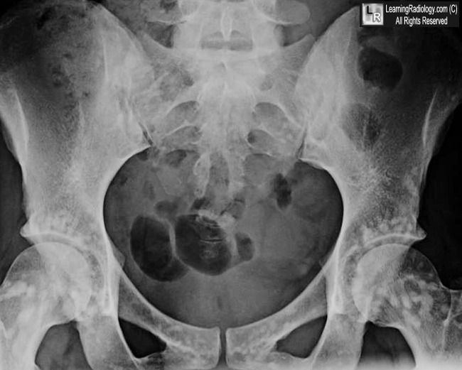
What is the most likely diagnosis?

  • 37 year-old female with abdominal pain


Frontal radiograph of the pelvis
  1. Osteoblastic breast metastases
  2. Dermoid of the ovary
  3. Osteopoikilosis
  4. Pelvic fracture
  5. Multiple myeloma
Additional Images - Axial CT scan of the pelvis


Recognizing
A Technically
Adequate Chest X-ray

Factors to Evaluate
lPenetration
lInspiration
lRotation
lAngulation


You should be able to
just see the thoracic
spine through the
heart.


Wednesday, August 11, 2010

Bronchiolitis

Bronchiolitis


Bronchiolitis
Classification and external resources

A chest X-ray demonstrating lung hyperinflation with a flattened diaphragm and bilateralatelectasis in the right apical and left basal regions in a 16-day-old infant with severe bronchiolitis.
ICD-10J21.
ICD-9466.1
DiseasesDB1701
MedlinePlus000975
eMedicineemerg/365
MeSHD001988
Bronchiolitis is inflammation of the bronchioles, the smallest air passages of the lungs. This inflammation is usually caused by viruses.

Contents

 [hide]

[edit]Causes

The term usually refers to acute viral bronchiolitis, a common disease in infancy. This is most commonly caused by respiratory syncytial virus (RSV, also known as human pneumovirus). (J21.0)
Other viruses which may cause this illness include metapneumovirus, influenza, parainfluenza,coronavirus, adenovirus, and rhinovirus.
The American Academy of Pediatrics has published a clinical practice guideline for theDiagnosis and Management of Bronchiolitis, including a review of the evidence and recommendations.

[edit]Diagnosis and Recovery

In a typical case, an infant under two years of age develops cough, wheeze, and shortness of breath over one or two days. The diagnosis is made by clinical examination. Chest X-ray is sometimes useful to exclude pneumonia, but not indicated in routine cases.
Testing for specific viral cause (e.g. RSV by nasopharyngeal aspirate) can be done but has little effect on management. In a systematic review, RSV testing by direct immunofluorescence testing on nasopharyngeal aspirate had a sensitivity of 61% and specificity of 89%.[1]Identification of RSV-positive patients can be helpful for:
  • disease surveillance
  • grouping ("cohorting") patients together in hospital wards as to prevent cross infection
  • predicting whether the disease course has peaked yet
  • reducing the need for other diagnostic procedures (by providing confidence that a cause has been identified).
The infant may be breathless for several days. After the acute illness, it is common for the airways to remain sensitive for several weeks, leading to recurrent cough and wheeze.
There is a possible link with later asthma: possible explanations are that bronchiolitis causes asthma by inducing long term inflammation, or that children who are destined to be asthmatic are more susceptible to develop bronchiolitis.
Bronchiolitis Obliterans occurs rarely in rheumatoid lung disease when small airway obstruction develops into a necrotizing bronchiolitis.

[edit]Treatment

There is no effective specific treatment for bronchiolitis. Therapy is principally supportive.[2]

[edit]Conservative measures

Frequent small feeds are encouraged to maintain hydration as evidenced by good urine output, and sometimes oxygen may be required to maintain blood oxygen levels. Suction of the nasopharynx is often performed to maintain a clear airway. In severe cases the infant may need to be fed via a nasogastric tube or it may even need intravenous fluids. In extreme cases, mechanical ventilation (for example, using CPAP) might be necessary.

[edit]Bronchodilators

Bronchodilator drugs such as salbutamol/albuterol or ipratropium are no longer recommended, but many clinicians offer a trial dose to see if there is any benefit (especially if there is a family history of asthma, since it can be difficult to clinically distinguish bronchiolitis from a viral-induced asthma). Racemic epinephrine is another drug that is sometimes given.

[edit]Hypertonic saline

There is some interest in the use of hypertonic saline in bronchiolitis. Initially recommended for use in cystic fibrosis patients, it is speculated to increase hydration of secretions, thus facilitating their removal. [3] In a study of emergency room patients with bronchiolitis, a comparison of nebulized 3% saline/epinephrine with 0.9% saline/epinephrine showed no difference in effectiveness.[4] However, in another study looking at the inpatient population, aerosolized 3% saline soln/1.5mg epinephrine was shown to reduce the length of hospital stay among non-asthmatic, non-severely ill infants with viral bronchiolitis as compared to aerosolized 0.9% saline soln/1.5mg epinephrine according to a randomized controlled trial. Length of stay for aerosolized 3% saline soln/1.5mg epinephrine was 3 +/- 1.2 days, which was significantly shorter (by 25%) than length of stay for patients treated with 0.9% saline soln/1.5mg epinephrine which was 4 +/- 1.9 days (p < 0.05).[5]

[edit]Non effective

Ribavirin is an antiviral drug which has a controversial role in treating RSV infection. There is no proven benefit but it is used sometimes for infants with pre-existing lung, heart or immune disease. Antibiotics are often given in case of a bacterial infection complicating bronchiolitis, but have no effect on the underlying viral infection.
Corticosteroids have no proven benefit in bronchiolitis treatment and are not advised.
DNAse has not been found to be effective.[6]

[edit]Complications

Middle ear bacterial infection
Development of asthma later (bronchial hyperactivity)

[edit]Prevention

In general, prevention of bronchiolitis relies on measures to reduce the spread of the viruses that cause respiratory infections (that is, handwashing, and avoiding exposure to those symptomatic with respiratory infections).
Premature infants, and others with certain major cardiac and respiratory disorders, can receive passive immunization with Palivizumab (amonoclonal antibody against RSV). This form of passive immunization therapy requires monthly injections every winter. Whether it could benefit infants with lung problems secondary to muscular dystrophies and other vulnerable groups is currently unknown


Introduction

Background

Bronchiolitis is an acute infectious disease of the lower respiratory tract that occurs primarily in young infants, most often in those aged 2-24 months.

Pathophysiology

Bronchiolitis is usually due to a viral infection of the small airways (bronchioles). Infection of bronchiolar respiratory and ciliated epithelial cells produces increased mucus secretion, cell death, and sloughing, followed by a peribronchiolar lymphocytic infiltrate and submucosal edema. The combination of debris and edema produces critical narrowing and obstruction of small airways.
Decreased ventilation of portions of the lung causes ventilation/perfusion mismatching, resulting in hypoxia. During the expiratory phase of respiration, further dynamic narrowing of the airways produces disproportionate airflow decrease and resultant air trapping. Work of breathing is increased due to increased end-expiratory lung volume and decreased lung compliance. Recovery of pulmonary epithelial cells occurs after 3-4 days, but cilia do not regenerate for about 2 weeks. The debris is cleared by macrophages.
Infection is spread by direct contact with respiratory secretions. In the United States, epidemics last 2-4 months beginning in November and peaking in January or February. While 93% of cases occur between November and early April, sporadic cases may occur throughout the year. Attack rates within families are as high as 45% and are higher in daycare centers. Rates of hospital-acquired infection range from 20-47%. Previous infection with the common etiologic viruses does not confer immunity. Reinfection is common.

Frequency

United States

Annual incidence is 11.4% in children younger than 1 year and 6% in those aged 1-2 years. The illness accounts for 4500 deaths and 90,000 hospital admissions per year. Prevalence may be higher in urban areas.
In children aged 2 years, approximately 95% have serologic evidence of past infection with the predominant causative agent, respiratory syncytial virus (RSV). Unfortunately, presence of antibodies to RSV does not confer immunity.

International

RSV is an important respiratory pathogen worldwide. The frequency of bronchiolitis in developed countries appears to be similar to that in the United States. Epidemiologic data for underdeveloped countries are incomplete. Peak incidence of bronchiolitis usually occurs during winter months in temperate climates and during the rainy season in tropical climates. In the United States, the peak occurs in February. Morbidity and mortality may be higher in less developed countries because of poor nutrition and lack of resources for supportive medical care.

Mortality/Morbidity

Significant morbidity is unusual.
  • Hospitalization is required in up to 2% of cases; most of those patients are younger than 6 months. These patients account for as many as 17% of all infant hospitalizations. Hospitalization is significantly more likely at altitudes above 2500 meters (8000 ft). Mechanical ventilation is required for 3-7% of admitted patients.
  • The mortality rate is 1-2% of all hospitalized patients and 3-4% for patients with underlying cardiac or pulmonary disease.
  • Most deaths occur in infants younger than 6 months.

Race

  • Race and socioeconomic status may affect the frequency of contracting bronchiolitis.
  • Lower socioeconomic status may increase the likelihood of hospitalization. Hospitalization rates are higher in Native American, Alaskan, and Hispanic populations, but it is not clear if this is due to more severe infection or a lower threshold for admission.

Sex

Bronchiolitis occurs as many as 1.25 times more frequently in males than in females.

Age

  • Although infection with etiologic agents may occur at any age, the clinical entity of bronchiolitis includes only infants and young children. Seventy-five percent of cases of bronchiolitis occur in children younger than 1 year, and 95% in children younger than 2 years. Incidence peaks in those aged 2-8 months.

Clinical

History

  • History and physical examination form the primary basis for the diagnosis of bronchiolitis.
  • Early symptoms are those of a viral upper respiratory tract infection (URI), including mild rhinorrhea, cough, and sometimes low-grade fever.
  • Adults, older children, and many infants do not progress beyond this stage of URI.
  • For the 40% of infants and young children who progress to lower respiratory tract involvement, paroxysmal cough and dyspnea develop within 1-2 days.
  • Other common symptoms include the following:
    • Fever
    • Increased work of breathing
    • Wheezing
    • Cyanosis
    • Grunting
    • Noisy breathing
    • Vomiting, especially post-tussive
    • Irritability
    • Poor feeding or anorexia

Physical

Most patients with bronchiolitis have the following signs:
  • Tachypnea, often at rates over 50-60 breaths per minute (most common physical sign)
  • Tachycardia
  • Fever, usually in the range of 38.5-39°C
  • Mild conjunctivitis or pharyngitis
  • Diffuse expiratory wheezing
  • Nasal flaring
  • Intercostal retractions
  • Cyanosis
  • Inspiratory crackles
  • Otitis media
  • Apnea, especially in infants younger than 6 weeks
  • Palpable liver and spleen from hyperinflation of the lungs and consequent depression of the diaphragm

Causes

RSV is the most common pathogen (85%), but other organisms occasionally produce a similar clinical picture.
  • Adenovirus (11%) occasionally causes a similar syndrome with a more virulent course.
  • Epidemics of bronchiolitis due to parainfluenza virus usually begin earlier in the year and tend to occur every other year.
  • Other less common etiologic agents include the following:
  • Ear, nose, or mouth inoculation
    • Exposure to an adult with a URI
    • Daycare exposure (significant)
    • Idiopathic


Hydronephrosis

Hydronephrosis is a condition in which the kidney swells, due to a backup of urine. Hydronephrosis generally occurs with another disease such as acute or chronic unilateral obstructive uropathy, vesicoureteric reflux, kidney stones, and during pregnancy. Symptoms of hydronephrosis include nausea, vomiting, urinary tract infection, fever, painful urination, increased urinary frequency and urgency, flank pain, and swelling of the abdomen. Treatment of hydronephrosis depends on the cause.


Hydronephrosis
Classification and external resources

Specimen of a kidney that has undergone extensive dilation due to hydronephrosis. Note the extensive atrophy and thinning of the renal cortex.
ICD-10N13.0-N13.3
ICD-9591
DiseasesDB6145
MeSHD006869
Hydronephrosis is distension and dilation of the renal pelvis and calyces, usually caused byobstruction of the free flow of urine from the kidney, leading to progressive atrophy of the kidney.[1] In case of hydroureteronephrosis, there is distention of both the ureter and the renal pelvis and calices.[2]

Contents

 [hide]

[edit]Signs and symptoms

The signs and symptoms of hydronephrosis depend upon whether the obstruction is acute orchronic, partial or complete, unilateral or bilateral. Unilateral hydronephrosis may occur without any symptoms, while acute obstruction can cause intense pain.[1]
Blood tests can show raised creatinine, urea and electrolyte imbalance. Urinalysis may show an elevated pH due to the secondary destruction of nephrons within the affected kidney.
Symptoms that occur regardless of where the obstruction lies include loin or flank pain. An enlarged kidney may be palpable on examination.
Where the obstruction occurs in the lower urinary tract, suprapubic tenderness (with or without a history of bladder outflow obstruction) along with a palpable bladder are strongly suggestive ofacute urinary retention, which left untreated is highly likely to cause hydronephrosis.
Upper urinary tract obstruction is characterised by pain in the flank, often radiating to either theabdomen or the groin. Where the obstruction is chronic, renal failure may also be present. If the obstruction is complete, an enlarged kidney is often palpable on examination.

[edit]Aetiology

The obstruction may be either partial or complete and can occur anywhere from the urethral meatus to the calyces of the renal pelvis.
The obstruction may arise from either inside or outside the urinary tract or may come from the wall of the urinary tract itself. Intrinsic obstructions (those that occur within the tract) include blood clots, stones, sloughed papilla along with tumors of the kidney, ureter and bladder. Extrinsic obstructions (those that are caused by factors outside of the urinary tract) include pelvic or abdominal tumours or masses, retroperitoneal fibrosis or neurological deficits. Strictures of the ureters (congenital or acquired), neuromuscular dysfunctions or schistosomiasis are other causes which originate from the wall of the urinary tract.
Hardikar syndrome can include Hydronephrosis, Cleft lip and palate, Vesicoureteral reflux, Intestinal obstruction, Obstructive liver disease and other symptoms. [3]

[edit]Tests

Ultrasound picture of hydronephrosis caused by a left ureteral stone.
Prenatal diagnosis is possible.[4]
Blood (U&E, creatinine) and urine (MSU, pH) tests should be taken. IVUs, ultrasounds, CTs and MRIs are also important tests. Ultrasound allows for visualisation of the ureters and kidneys and can be used to assess the presence of hydronephrosis and / or hydroureter. An IVU is useful for assessing the position of the obstruction. Antegrade or retrograde pyelography will show similar findings to an IVU but offer a therapeutic option as well.
The choice of imaging depends on the clinical presentation (history, symptoms and examination findings). In the case of renal colic (one sided loin pain usually accompanied by a trace of blood in the urine) the initial investigation is usually a spiral or helical CT scan. This has the advantage of showing whether there is any obstruction of flow of urine causing hydronephrosis as well as demonstrating the function of the other kidney. Many stones are not visible on plain X-ray or IVU but 99% of stones are visible on CT and therefore CT is becoming a common choice of initial investigation. CT is not used however, when there is a reason to avoid radiation exposure, e.g. in pregnancy.

[edit]Prognosis

Left untreated, bilateral obstruction (occurring to both kidneys rather than one) has a poor prognosis.

[edit]Treatment

Treatment of hydronephrosis focuses upon the removal of the obstruction and drainage of the urine that has accumulated behind the obstruction. Therefore, the specific treatment depends upon where the obstruction lies, and whether it is acute or chronic.
Acute obstruction of the upper urinary tract is usually treated by the insertion of a nephrostomy tube. Chronic upper urinary tract obstruction is treated by the insertion of a ureteric stent or a pyeloplasty.
Lower urinary tract obstruction (such as that caused by bladder outflow obstruction secondary to prostatic hypertrophy) is usually treated by insertion of a urinary catheter or a suprapubic catheter.
Surgery is not required in all cases


Hydronephrosis
Pronunciations
Hydronephrosis is distention (dilation) of the kidney with urine, caused by backward pressure on the kidney when the flow of urine is obstructed.
  • Kidney stones are common causes of urinary tract obstruction.
  • When hydronephrosis occurs quickly, people may have excruciating pain, most often in the flank (the area between the ribs and the hips).
  • When hydronephrosis occurs more gradually, people may have no symptoms or experience attacks of dull, aching discomfort in the flank.
  • Doctors initially use bladder catheterization (or ultrasonography) to detect hydronephrosis, and they may use ultrasonography or another imaging test to determine the site of the blockage.
  • Treatment depends on the cause of the obstruction.
Hydronephrosis: A Distended Kidney
In hydronephrosis, the kidney is distended because the flow of urine is obstructed and urine backs up in the kidney's small tubes and central collecting area (renal pelvis).
Normally, urine flows out of the kidneys at extremely low pressure. If the flow of urine is obstructed, urine backs up behind the point of blockage, eventually reaching the small tubes of the kidney and its collecting area (renal pelvis), distending the kidney and increasing the pressure on its internal structures. The elevated pressure from obstruction may ultimately damage the kidney and can result in loss of its function. When the flow of urine is obstructed, urinary tract infections are fairly common and stones are more likely to form. If both kidneys are obstructed, kidney failure may result.
Long-standing distention of the renal pelvis and ureter can also inhibit the rhythmic muscular contractions that normally move urine down the ureter from the kidney to the bladder (peristalsis). Scar tissue may then replace the normal muscular tissue in the walls of the ureter, resulting in permanent damage.
Causes
Hydronephrosis commonly results from an obstruction located at the junction of the ureter and renal pelvis (ureteropelvic junction). Causes of this type of obstruction include the following:
  • Structural abnormalities—for example, a birth defect in which the insertion of the ureter into the renal pelvis is too high or there is inadequate development of the ureteral muscles (congenital ureteropelvic junction obstruction)
  • Kinking at the ureteropelvic junction resulting from a kidney shifting downward (ptosis of the kidney)
  • Stones (calculi) or a blood clot in the renal pelvis
  • Compression of the ureter by bands of fibrous tissue, an abnormally located artery or vein, or a tumor
Hydronephrosis can also result from an obstruction below the ureteropelvic junction or from backflow (reflux) of urine from the bladder. Causes of this type of obstruction include the following:
  • Stones in the ureter
  • Blood clot in the ureter
  • Tumors in or near the ureter
  • Narrowing of the ureter resulting from a birth defect, an injury, an infection, radiation therapy, or surgery
  • Disorders of the muscles or nerves in the ureter or bladder
  • Formation of fibrous tissue in or around the ureter resulting from surgery, radiation therapy, or drugs (especially methysergide)
  • Bulging of the lower end of the ureter into the bladder (ureterocele)
  • Cancers of the bladder, cervix, uterus, prostate, or other pelvic organs
  • Obstruction that prevents urine flow from the bladder to the urethra, resulting from prostate enlargement (most often caused by a condition called benign prostatic hyperplasia—see Prostate Disorders: Benign Prostatic Hyperplasia (BPH)), or rectal impaction with feces
  • Abnormal contractions of the bladder resulting from a birth defect or a spinal cord or nerve injury
Hydronephrosis of both kidneys can occur during pregnancy as the enlarging uterus compresses the ureters. Hormonal changes during pregnancy may aggravate the problem by reducing the muscular contractions that normally move urine down the ureters. This condition, commonly called hydronephrosis of pregnancy, usually ends when the pregnancy ends, although the renal pelvis and ureters may remain somewhat distended afterward.
Symptoms
Symptoms depend on the cause, location, and duration of the obstruction. When the obstruction begins quickly (acute hydronephrosis), it usually produces renal colic—an excruciating, intermittent pain in the flank (the area between the ribs and hip) on the affected side. Obstruction on one side does not reduce urine flow. Obstruction can stop or reduce urine flow if blockage affects the ureters from both kidneys or if it affects the urethra. Obstruction of the urethra or bladder outlet may produce pain, pressure, and distention of the bladder.
People who have slowly progressive (chronic) hydronephrosis may have no symptoms, or they may have attacks of dull, aching discomfort in the flank on the affected side. Sometimes a kidney stone temporarily blocks the ureter and produces painful hydronephrosis that occurs intermittently.
Hydronephrosis may cause vague intestinal symptoms, such as nausea, vomiting, and abdominal pain. These symptoms sometimes occur in children when hydronephrosis results from a birth defect in which the junction of the ureter and renal pelvis is too narrow (ureteropelvic junction obstruction).
People who have urinary tract infections may have pus in the urine, fever, and discomfort in the area of the bladder or kidneys.
Diagnosis
Early diagnosis is important, because most cases of obstruction can be corrected and because a delay in treatment can lead to irreversible kidney damage. Doctors may suspect hydronephrosis because of a person's symptoms and sometimes because of findings discovered during a physical examination. A distended kidney can occasionally be felt in the flank, particularly if the kidney is greatly enlarged in an infant or a child or a thin adult. A distended bladder can sometimes be felt in the lower part of the abdomen just above the pubic bone.
Doctors depend on testing to make the diagnosis. Bladder catheterization (insertion of a hollow, flexible tube through the urethra) is often the first diagnostic test done in people with renal colic, pelvic pressure, or distention. If the catheter drains a large amount of urine from the bladder, then either the bladder outlet or the urethra is obstructed. Many doctors do ultrasonography to determine whether the bladder is filled with a large amount of urine before doing bladder catheterization.
If the presence or site of obstruction is in doubt, various imaging tests can be done to identify evidence of obstruction such as hydronephrosis or a site of blockage. For example, ultrasonography is a very useful test in most people (particularly children and pregnant women) because it is fairly accurate and does not expose the person to any radiation. Computed tomography (CT) scanning is an alternative. It is rapid and highly accurate, particularly at identifying stones. Other imaging tests, such as intravenous urography, may be performed to identify the site of obstruction, if it is not visible with ultrasonography or CT.
An endoscope (a rigid or flexible telescope) is sometimes used to look at possible sites of obstruction as closely as possible. An endoscope can be used to examine the urinary tract.
Blood and urine tests are done. Blood test results are usually normal, but tests may reveal high levels of urea nitrogen (sometimes called BUN), creatinine, or both, if obstruction affects both kidneys. Results from an analysis of urine (urinalysis) are usually normal but white blood cells and red blood cells may be present when a stone or a cancer is the cause of obstruction, or when the obstruction is complicated by an infection.
Prognosis
Permanent kidney damage is unlikely to result unless both kidneys are obstructed for at least a few weeks. The prognosis is less certain for chronic hydronephrosis.
Treatment
Treatment usually aims to relieve the cause of obstruction. For example, if the urethra is obstructed because of an enlarged or cancerous prostate, treatment can include drugs, such as hormone therapy for prostate cancer (see Prostate Disorders: Prostate Cancer), surgery, or enlargement of the urethra with dilators. Other treatments, such as lithotripsy or endoscopic surgery, may be needed for stones that block the flow of urine. If the cause of obstruction cannot be rapidly corrected, particularly if there is infection, kidney failure, or severe pain, the urinary tract is drained. In acute hydronephrosis, urine that has accumulated above the obstruction can be drained with a soft tube inserted through the skin into the kidney (nephrostomy tube) or by insertion of a soft plastic tube that connects the bladder with the kidney (ureteral stent). Complications of nephrostomy tubes or ureteral stents can include displacement of the tube, infection, and discomfort.
Urgent relief of chronic hydronephrosis is usually not required. Complications of hydronephrosis, such as urinary tract infections and kidney failure, if present, are treated promptly.
Last full review/revision August 2007 by Glenn M. Preminger, MD